Synonyms: Antibodies to thyroid peroxidase, AT-TPO, microsomal antibodies, anti-thyroid, antibodies to microsomal antigen, ATPO.
Scientific editor: M. Merkusheva, PSPbSMU named after. acad. Pavlova, medical practice. Proofreader: M. Mazur, KSMU named after. S. I. Georgievsky, therapist
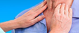
The thyroid gland plays a responsible role in the body - the production of biologically active substances that are responsible for the process of energy exchange between cells. Their secretion occurs with the participation of a special enzyme - thyroid peroxidase (TPO), it is involved in the formation of the active form of iodine, without which the biochemical synthesis of thyroid hormones T4 and T3 is impossible.
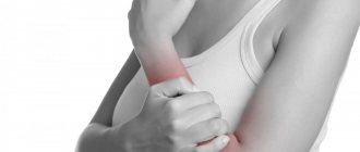
Antibodies (AT) to TPO are formed when the enzyme is detected by the body as a foreign protein. The AT-TPO analysis is a highly accurate marker that determines the level of aggression of the immune system towards its own body, and allows for timely diagnosis of autoimmune diseases of the thyroid gland: diffuse toxic goiter, thyroiditis, thyroid dysfunction in infants.
General information
To thyroid peroxidase, which is located on the surface of thyrocytes (cells that produce T3 and T4), i.e. directly in the thyroid gland, the immune system does not react in any way. But only up to a certain point. When this enzyme enters the blood, and this happens in the case of damage to the thyroid gland, provoked by external or internal factors, the active synthesis of autoantibodies to peroxidase (AT-TPO) begins in the body.
Provoking factors:
- radiation therapy (in the treatment of cancer), systematic irradiation of the body (occupational hazard);
- trauma to the thyroid gland as a result of a bruise, blow, fall, puncture, etc.;
- unsuccessful surgical intervention on the gland;
- deficiency or excess of iodine in the body;
- inflammatory processes, infectious and viral diseases.
When the amount of antibodies increases, massive destruction of peroxidase and thyroid follicular cells secreting T3 and T4 begins. As a result, the concentration of these hormones in the blood increases sharply. This condition is diagnosed as autoimmune thyrotoxicosis. Then, over the course of 1.5-2 months, T3 and T4 are washed out of the body, and their levels in the blood drop. At the same time, there is no possibility of replenishing the hormone deficiency, because the cells that produce them are completely destroyed. Hypothyroidism develops.
If the amount of AT has increased moderately, then over the course of decades they will gradually destroy thyroid cells and gradually reduce the volume of hormones produced. As a result, the patient will develop insufficient thyroid function, and there will be a deficiency of the most important iodinated hormones (T3 and T4). This is the same hypothyroidism.
The AT-TPO test makes it possible to accurately diagnose pathological conditions, the correction of which requires the use of hormone replacement therapy (HRT). With the correct dosage of synthetic hormones (levothyroxine), such treatment tactics provide a stable and long-lasting clinical effect.
To improve your appearance
Support for Turbo pages on a wider class of mobile screens - so that your Turbo pages look equally good not only on mobile, but also on tablets.
Transferring html layout directly to RSS. To make it more convenient for you to work with the appearance of Turbo pages, we will add the ability to transfer your own html layout directly to RSS. It will be possible to transfer ready-made blocks of your website to the Turbo page practically without changes and not waste time re-configuring the appearance.
Settings for basic design elements without CSS. And so that you can save time on developing your own CSS, let's add the following features:
- customize color, font, logo and other elements directly in Webmaster,
- quickly enable light and dark themes on your website.
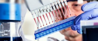
Indications for analysis
In addition to direct indications (diagnosis of autoimmune thyroid diseases), an endocrinologist may prescribe an anti-TPO antibody test in the following cases:
- when the patient complains of: uncontrolled weight gain or loss, chronic fatigue, constantly low or, conversely, increased body temperature, increased sweating, increased anxiety, insomnia.
- if the results of other tests (T3, T4 and/or TSH) indicate thyroid dysfunction.
- determining the risk of neonatal hypothyroidism (thyroid deficiency in newborns), if the mother has a history of thyroid disease or antibodies to TPO are detected;
- screening in the first trimester of pregnancy to determine the risk of developing thyroiditis (inflammation of the thyroid gland);
- screening in pregnant women with TSH (thyroid-stimulating hormone) concentration >2.5;
- determination of the degree of risk of miscarriage, spontaneous termination of pregnancy (miscarriage);
- diagnosis of female infertility and unsuccessful attempts at artificial insemination;
- assessment of the structure and condition of the thyroid gland before prescribing medications (HRT, amiodarone, interferon, lithium or iodine preparations, etc.);
- diagnosis of hypothyroidism, goiter (enlargement of the gland), thyroiditis, thyrotoxicosis (excessive secretion of iodinated hormones);
- clarification of ultrasound results that revealed a disorder of the structure (heterogeneity) of the thyroid gland;
- to examine the thyroid gland in newborns to ensure that there are no abnormalities if the mother has been diagnosed with antibodies to thyroid peroxidase or postpartum thyroiditis.
Thyroiditis in children
The thyroid gland is located on the front surface of the neck and, more than other endocrine organs, is exposed to the influence of the external environment, and the unity of the lymphatic ring of the pharyngeal tonsils and the thyroid gland creates conditions for the occurrence of an inflammatory process in it against the background of chronic diseases of the nasopharynx.
The child's body is predisposed to the development of autoimmune processes, since its lymphatic system is more active than that of adults.
Most often, existing classifications of inflammatory diseases of the thyroid gland are based on external symptoms. In particular, our classification includes: a) acute thyroiditis (purulent and non-purulent); b) subacute thyroiditis; c) chronic thyroiditis (fibrous - Riedel and lymphomatous - Hashimoto); d) thyroid tuberculosis; e) syphilis of the thyroid gland; f) parasitic diseases; g) actinomycosis of the thyroid gland. Obviously, the concept of “inflammatory diseases of the thyroid gland” includes pathological conditions that have different pathogenetic essences, in particular, thyroiditis often has an autoimmune rather than inflammatory genesis. This approach to the classification of this pathology still causes controversy among specialists.
Inflammatory diseases of the thyroid gland are relatively rare (1–2% of all thyroid diseases). Thyroiditis is an inflammation of a previously normal thyroid gland (strumitis is an inflammatory process in a goitrous gland). Thyroiditis is distinguished between nonspecific (acute, subacute and chronic) and specific - tuberculous, syphilitic, actinomycotic, parasitic.
Acute thyroiditis
The cause of the disease can be any acute or chronic infection (flu, tonsillitis, acute respiratory infections, chronic tonsillitis, scarlet fever, mumps, tuberculosis, syphilis, etc.).
Inflammation can develop either directly during infection or as a complication. The cause of its occurrence can also be various intoxications with iodine, lead, as well as traumatic damage to the capsule. In some cases, the etiology of thyroiditis cannot be established.
Clinical manifestations of acute thyroiditis are variable. More often the process begins unnoticed, gradually. There is pain in the neck, sometimes pain when swallowing, slight hoarseness, weakness, and malaise. The temperature reaction is often absent; less often, an increase in temperature may be observed. Often during this period the disease remains unattended, and the doctor’s efforts are aimed at examining the nasopharynx.
After a few days (sometimes after 2–3 months), the temperature rises, chills, weakness, headache, sweating, tachycardia, and in some cases nausea and vomiting appear. The thyroid gland increases in size, becomes dense and sharply painful. Movement of the head, coughing, and swallowing increase pain, which can radiate to the head, ears, and jaw. Swelling and dilation of the veins of the neck, pastiness and hyperemia of the face are noted, the cervical lymph nodes increase in size, moderate leukocytosis with a shift to the left, and accelerated ESR are detected in the blood.
At the onset of the disease, there are signs of increased functional activity of the thyroid gland.
In the future, signs of hypothyroidism are sometimes detected, which are also transient in most cases.
The acute period lasts an average of 3–6 weeks, after which the temperature normalizes, pain disappears, and the size of the thyroid gland decreases. Due to the appearance of the inflammatory process in other parts of the gland, relapses of the disease may occur, so recovery from thyroiditis (with restoration of thyroid function) can be said after 4–6 months from the onset of the disease.
Along with cases of thyroiditis that spontaneously end in recovery, severe, fulminant forms may occur, which are accompanied by necrosis and purulent melting of the parenchyma. These forms are often caused by streptococci, staphylococci, and pneumococci. In these cases, the disease develops acutely and is characterized by a general severe condition, a significant increase in temperature, chills, tachycardia, severe pain in the anterior surface of the neck, and often a feeling of suffocation. There is significant leukocytosis (15,000–20,000), neutrophilia, a shift in the leukocyte formula to the left, and accelerated ESR.
Suppuration can be diffuse and localized in the form of an abscess. A breakthrough of pus occurs through the skin, as well as the esophagus, larynx or trachea (aspiration pneumonia), and mediastinum (mediastenitis).
Purulent thyroiditis, more often than other forms, ends with the development of fibrosis of the thyroid gland, which leads to a decrease in its functional activity and the development of hypothyroidism.
Treatment of acute thyroiditis should include dietary nutrition (liquid, semi-liquid food in small portions). The patient is prescribed bed rest during the entire acute period, the position in bed is semi-sitting.
Antibiotic therapy is mandatory, in large doses during the first 7–10 days. The average course of antibiotic treatment is 3–4 weeks.
At the same time, desensitizing agents are prescribed in age-specific doses throughout the entire acute period. According to indications, oxygen therapy, cardiac, painkillers, and sleeping pills are used. For hypofunction of the thyroid gland, treatment is carried out with L-thyroxine (50–100 mcg). It is advisable to prescribe vitamin therapy (A, C, B1, B2, B12).
Warming compresses, diathermy, and Solux are not indicated for everyone.
Treatment of acute thyroiditis should begin as early as possible, the doses of drugs should be large enough, and treatment should not be stopped immediately after the signs of inflammation are eliminated.
For purulent thyroiditis, surgical treatment (opening of the abscess and drainage) is indicated.
Subacute thyroiditis
Subacute thyroiditis, known as giant cell granulomatous thyroiditis, is a rare disease. Recently, it has been assumed that it is viral in nature. Often the disease develops after suffering from influenza, measles, mumps and other infections. In a significant proportion of patients, autoantibodies to thyroid tissues are detected in the blood serum, which suggests the presence of autoimmune mechanisms in the pathogenesis of this disease. Subacute thyroiditis is characterized by the formation of giant cells and granulomas resembling a tuberculous tubercle, as well as lymphoid-plasmacytic infiltration of the stroma, which is characteristic of an autoimmune process.
Clinically, subacute thyroiditis is characterized by pain in the thyroid gland, radiating to the parotid region, neck, and back of the head. The thyroid gland increases in size and is painful on palpation. Symptoms of intoxication are noted: weakness, lethargy, fever.
The disease is often accompanied by dysfunction of the thyroid gland, with the appearance of symptoms of thyrotoxicosis: weakness, sweating, irritability, tachycardia. Subsequently, the phase of hyperthyroidism is replaced by symptoms of hypothyroidism, due to depletion of the synthesis of thyroid hormones. The levels of triiodothyronine (T3) and thyroxine (T4) in the blood decrease, and the content of thyroid-stimulating hormone increases.
At the peak of the disease, accelerated ESR, dysproteinemia (increased α2- and γ-globulin fractions) are detected, and antibodies to thyroglobulin in low titers are detected in the blood.
In the treatment of these forms of thyroiditis, the use of glucocorticoids is of particular importance. Salicylic or pyrazolone drugs are also used. Antibiotics are prescribed for the period of hormonal therapy and have no independent value.
Prescribing methizol in the hyperthyroid phase is not recommended, since thyrotoxicosis is caused by the destruction of thyrocytes and the release of previously synthesized hormones into the blood. It is more advisable to use β-blockers.
Considering that hypothyroidism in subacute thyroiditis is reversible, taking thyroid hormones is not required. Normalization of the echostructure of the thyroid gland in patients occurs with a delay compared to the normalization of clinical and laboratory parameters.
Chronic fibrous thyroiditis
Chronic fibrous thyroiditis (Riedel's goiter) is rare.
Microscopically, the thyroid gland reveals a chronic productive inflammatory nonspecific process with a pronounced proliferation of dense fibrous connective tissue with a certain amount of diffuse lymphoid cell infiltrates and very often with the presence of eosinophils, which is one of the characteristic signs of this disease. The architecture of the thyroid gland is disturbed.
The disease begins unnoticed, a protrusion appears on the neck, which is not accompanied by pain. When the trachea and esophagus are involved in the process, breathing and swallowing disorders, hoarseness, and dry cough are noted.
The thyroid gland is often diffusely enlarged. The consistency is very dense, woody, the surface is smooth. The skin over the gland is not changed. Regional lymph nodes are not enlarged. With a long course of the disease, adhesion of the goiter to the surrounding tissues is noted, which may suggest the presence of a malignant tumor of the thyroid gland. The general condition, as a rule, is not disturbed, the function of the thyroid gland does not change.
An X-ray examination reveals signs of displacement or compression of the trachea and esophagus.
The generally accepted method is surgical treatment of this disease, which consists of resection of the isthmus and nearby areas of the lobes to relieve the symptoms of tracheal compression. After the operation, the process may reverse.
Chronic autoimmune thyroiditis
Among inflammatory diseases of the thyroid gland in children, a large proportion belongs to chronic autoimmune thyroiditis (CHAT). This is due to the deterioration of the environmental situation, as well as the unity of the lymphatic ring of the pharyngeal tonsils and the thyroid gland, which creates conditions for the occurrence of an inflammatory process in it against the background of chronic tonsillitis.
Autoimmune damage to the thyroid gland was first described by Hashimoto in 1912. The author called this disease Struma lymphomatosa.
With the accumulation of clinical data related to autoimmune damage to the thyroid gland, clinical classifications have been developed, the most successful of which is the classification of R. Volpe (1984), in the rubric of which ChAT is divided into several different forms, among which lymphocytic thyroiditis of children and adolescents is specifically distinguished.
In the pathogenesis of CHAT, the main role is played by disorders of immunological control in hereditarily predisposed children. It is known that ChAT according to the HLA system is associated with the DR 3 and DR 5 loci, like many other autoimmune diseases, and the association with the DR 5 locus causes the hypertrophic, and with the DR 3 locus, the atrophic form of ChAT.
The hereditary genesis of the disease is confirmed by data on frequent cases of the disease in close relatives. Other autoimmune diseases of endocrine and somatic origin are often recorded in the pedigree of patients with CAT, which may indicate an innate predisposition to autoimmune reactions.
A genetically determined defect in immunocompetent cells leads to a breakdown of natural tolerance and activation of cellular and humoral immunity. This process can be caused by a viral infection, medications, or, less commonly, injury. However, there is still no convincing data on the etiology of CHAT.
The process of producing antibodies to thyroglobulin, the microsomal fraction of the thyroid gland, the thyroid-stimulating hormone receptor of the pituitary gland, the release of lymphotoxins, tumor necrosis factor, etc. is launched, which ultimately causes destructive changes in thyrocytes and a decrease in thyroid function. The outcome of ChAT is the development of hypothyroidism. This condition leads to an increased release of thyroid-stimulating hormone from the pituitary gland, which provokes the growth of surviving thyrocytes followed by their infiltration by lymphocytes, macrophages, and plasma cells.
Histological examination of the thyroid gland reveals diffuse or focal infiltration of lymphocytes and plasma cells. The presence of large epithelioid Hürthle-Ashkenazi cells is characteristic. Depending on the phase and duration of the process, stromal fibrosis can be detected. The disease occurs in hypertrophic and atrophic variants.
In children, CHAT often develops in the pre- and puberty period, less often in preschool and primary school age. In the vast majority of children, autoimmune thyroiditis is accompanied by the development of goiter; in 5–10% of patients, an atrophic form of the disease is noted.
ChAT may be one of the components of autoimmune polyendocrine syndrome (APS). For the first time, the combination of autoimmune thyroiditis with primary adrenal insufficiency was described by E. V. Schmidt. Currently, two types of APS are distinguished.
The first type of APS involves adrenal insufficiency, hypoparathyroidism, autoimmune thyroiditis, type 1 diabetes mellitus, candidiasis, nail dystrophy, hypoplasia of tooth enamel, vitiligo, alopecia, chronic active hepatitis, autoimmune gastritis, hypogonadism, malabsorption syndrome. This syndrome is caused by a mutation in the autoimmune regulator gene. The second type of APS includes type 1 diabetes mellitus, adrenal insufficiency, autoimmune thyroiditis, and hypogonadism.
ChAT can be combined with rheumatoid arthritis, pernicious anemia, and systemic lupus erythematosus.
The disease is 4–5 times more common in girls. A family predisposition for thyroid pathology is detected in 65% of children, and 3 times more often on the maternal side.
In most cases, CHAT develops gradually. Clinical diagnosis of CHAT in children is based on the syndrome of enlarged thyroid gland.
Currently, one should use a classification of goiter sizes based on palpation data (WHO, 1994). According to WHO criteria, the following sizes of the thyroid gland are distinguished:
- 0 degree - no goiter;
- I degree - the size of the lobes of the thyroid gland is larger than the distal phalanx of the thumb of the patient being examined, but is not visible to the eye;
- II degree - the goiter is palpable and visible to the eye.
In case of CAT in children, the size of the goiter most often corresponds to the II degree of increase in the lobes of the thyroid gland. On palpation, the thyroid gland is compacted, less often - elastic in consistency, heterogeneous, with an uneven surface, mobile when swallowing.
The chronic course of the disease ultimately leads to hypothyroidism. But unlike adults, 1/3 of patients with ChAT debut with symptoms of increased thyroid function. The hyperthyroid phase of ChAT - this phenomenon is called hashitoxicosis, in contrast to similar manifestations in diffuse toxic goiter - develops as a result of the destruction of thyroid tissue and the release of a large amount of deposited hormones into the blood. In these cases, against the background of an enlarged thyroid gland, tachycardia, changes in blood pressure with an increase in pulse pressure, tremor of the eyelids and outstretched fingers, and increased bowel movements are observed. As a rule, exophthalmos, one of the typical symptoms of diffuse toxic goiter, is not recorded at this phase of ChAT.
The hyperthyroid phase in its natural course lasts no more than six months. In the future, spontaneous remission may occur. In 19% of patients, symptoms of hypofunction of the thyroid gland are detected, which manifest themselves in decreased performance at school, a tendency to constipation, chilliness, bradycardia, and bradypsychism syndrome.
When studying thyroid hormones, despite various clinical dysfunctions of the thyroid gland, the levels of T4 and T3 remain within the normal range. With hypofunction of the thyroid gland, the levels of thyroid-stimulating hormone of the pituitary gland are 2–2.5 times higher than its average values.
In tests of children who are diagnosed with thyrotoxicosis upon admission to the hospital, high levels of thyroid hormones are detected, thyroid-stimulating hormone levels are at the lower limit of normal or are below normal values.
In case of CAT, ultrasound examination reveals a diffuse or focal decrease in the echodensity of thyroid tissue. Hypoechoic areas without clear contours are observed in most patients. In some patients, hyperechoic “strands”—foci of fibrosis—are visualized. In cases where the echographic picture resembles a nodular formation with unclear contours, a dynamic examination of such a gland is necessary.
Thyroid antibodies to thyroglobulin and thyroid peroxidase are detected in 80–95% of patients with CAT in various diagnostic titers.
Fine needle biopsy is the most accurate diagnostic method. The morphological sign of ChAT is typical lymphoid infiltration, which always predominates over the epithelial component. In addition to lymphocytes, the presence of plasma cells and macrophages, large Hürthle-Ashkenazi cells is characteristic. The main indication for a puncture biopsy is suspicion of thyroid nodules or cancer suspicion.
So, the disease does not have a specific clinical picture, just as none of the research methods by itself can reliably make a diagnosis of ChAT. A comprehensive assessment of the pathological process in the thyroid gland involves studying the echostructure, thyroid status, and thyroid antibodies.
In differential diagnosis, certain difficulties are presented by the hyperthyroid phase of ChAT and the presence of diffuse toxic goiter, since ultrasound data, thyroid antibodies, and the morphological picture of punctate do not always allow an accurate diagnosis.
To diagnose CHAT, one should rely on the following indicators.
- Typically, thyrotoxicosis develops at the onset of the disease, followed by restoration of thyroid function after 4–6 months or the gradual development of hypothyroidism.
- Symptoms of thyrotoxicosis do not reach the intensity that is observed with diffuse toxic goiter.
- Thyroid-stimulating antibodies are markers of diffuse toxic goiter, and their detection in patients with CAT indicates a combination of two autoimmune diseases.
- The presence of ChAT may be indicated by the rapid development of euthyroidism during treatment of thyrotoxicosis. The course dose of methizol in children with ChAT in our observations averaged 600 mg, which is lower than for Graves' disease.
Effective therapy for ChAT as an immunopathological disease has not been developed. In case of pain in the thyroid gland during palpation and spontaneous pain in it, non-steroidal anti-inflammatory drugs (ortofen, indomethacin) should be prescribed in an age-specific dosage.
There is no convincing data on the effectiveness of immunocorrective drugs. In the hyperthyroid phase of mild CHAT, it is advisable to start treatment with β-blockers and sedatives. For more severe thyrotoxicosis, treatment is carried out with methizol - 10–15 mg per day.
To treat hypothyroidism, L-thyroxine is prescribed at a dose of 50–100 mcg per day under the control of thyroid-stimulating hormone and T4.
Surgical intervention for ChAT is carried out when the goiter is large and in the case of a combination of ChAT with a tumor process in the thyroid tissue.
V. V. Smirnov , Doctor of Medical Sciences, Professor I. S. Mavricheva , Candidate of Medical Sciences, Russian State Medical University, Moscow
AT-TPO test for pregnant women
Sometimes an increase in AT-TPO occurs during pregnancy, during the restructuring of the immune system for bearing a fetus. At this time, the thyroid gland becomes more active, producing more hormones. This is sometimes regarded by the immune system as a disorder and promotes the synthesis of antibodies to TPO.
Testing for antibodies to thyroid peroxidase during pregnancy is carried out for expectant mothers for preventive purposes. If the concentration of antibodies to TPO is high, then the risk of developing postpartum thyroiditis increases by 50%.
According to statistics, pathology develops in 5-10% of women after childbirth. The thyroid gland is gradually destroyed under the influence of AT, after which thyrotoxicosis develops (oversaturation of the body with iodinated hormones). Sometimes the function of the gland is restored on its own, but 1/3 of patients may develop hypothyroidism - chronic hormone deficiency, which requires systematic use of hormonal medications.
If the level of other thyroid hormones (T3, T4, TSH) is elevated in the first trimester, then the endocrinologist prescribes an AT-TPO test.
During pregnancy, antibodies to TPO can significantly affect the development of the thyroid gland of the unborn child, since they are able to penetrate the placental barrier from the mother’s blood into the fetus’s body and cause neonatal hypothyroidism.
Important! The interpretation of the results is always carried out comprehensively. It is impossible to make an accurate diagnosis based on only one analysis.
conclusions
If a person has been tested for antibodies to thyroid peroxidase, and the required norm is exceeded, under no circumstances should you immediately panic. Small deviations in values are quite possible even among healthy people. If you still have some minor deviations, you can bring your tests back to normal without using additional medications. To do this, you will simply need to reconsider your diet and give up all sorts of bad habits, after first getting rid of excess weight. Most doctors advise completely avoiding regularly wearing necklaces and chains around the neck, as some metals can negatively affect the functioning of the thyroid gland.
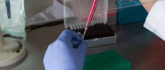
Standard for AT-TPO
All endocrinological studies (instrumental and laboratory) must be carried out in the same medical institution, since the reference values of AT-TPO in different laboratories may be different.
- According to the independent laboratory Invitro, for antibodies to thyroid peroxidase the indicator is set to 5.6 U/ml.
Standard reference values
The generally accepted norm (including abroad) is the indicator
- 0-34 IU/ml
However, there are also extended reference intervals:
Women:
- up to 50 years – 0-34 IU/ml.;
- after 50 years – 0-100 IU/ml;
Pregnant:
- at the 12th week of pregnancy – no higher than 25 IU/ml;
- in the 2nd and 3rd trimester - from 30 IU/ml to 56 IU/ml;
Men:
- up to 50 years - 0-34 IU/ml;
- after 50 years - less than 85 IU/ml.
Additional medical and public assistance
The importance of the thyroid gland should never be underestimated. And if malfunctions occur in its working functions, you need to immediately contact a doctor. As a rule, this refers to situations where there is a significantly increased titer of antibodies to an enzyme such as peroxidase. Treatment of this type of disorder is carried out through the use of medications. The doctor usually prescribes hormone replacement therapy on an individual basis.
As part of the development of autoimmune thyroiditis, the occurrence of hypothyroidism cannot usually be excluded. It is necessary to use medications until it becomes clear which one is most suitable.
For ordinary patients, just like pregnant women, doctors prescribe thyroid medications, for example, L-thyroxine. Patients are required to donate blood regularly. This is done so that the doctor can better examine the overall clinical picture and determine whether the treatment is successful.
AT-TPO is higher than normal
A significant deviation of the results from the norm is typical for:
- diffuse toxic goiter (Graves disease);
- nodular toxic goiter;
- thyroiditis (autoimmune, subacute (de Crevin's disease), chronic (Hashimoto's disease));
- postpartum thyroid dysfunction;
- idiopathic hypothyroidism;
A slight or moderate increase in AT-TPO levels may indicate the presence of:
- diabetes mellitus (insulin-dependent);
- rheumatoid arthritis (damage to joints and connective tissue);
- lupus erythematosus (an autoimmune pathology that affects connective tissue and skin);
- vasculitis (damage to vascular walls) and other autoimmune diseases.
Increase in AT-TPO during pregnancy
- Positive test results during pregnancy indicate the possibility of hyperthyroidism in the child (during intrauterine development or after birth)
The following factors can cause a false increase in AT-TPO:
- hereditary predisposition;
- a course of treatment with iodine or other drugs;
- chronic diseases in the acute stage;
- trauma or surgery in the thyroid gland.
For reference: about 5% of the world's population suffers from autoimmune thyroid diseases. This is about 350 million patients. In 10% of the remaining antibodies to TPO can be elevated without affecting the gland or caused by other systemic and autoimmune processes.
special instructions
But no matter how good and useful traditional medicine is, it is necessary to take into account that against the background of severely advanced forms of the disease, when AT to TPO is greatly increased (we explained what this means above), no herbs and herbal mixtures can correct the situation. Therefore, to prevent the patient’s condition from worsening further, it is necessary to engage in regular prevention. In addition, it is important to strictly observe and fulfill all medical instructions. Any signs that demonstrate or hint at problems in the proper functioning of the thyroid gland should be a signal and an incentive to urgently undergo the necessary examination to determine the causes of the disorders.