The ENT organs, being an outpost of the immune system on the path of infection into the body, are the first to encounter the effects of various pathogens. That is why inflammatory processes often begin in them. Inflammation of the paranasal sinuses is called sinusitis . In total, a person has 4 pairs of paranasal sinuses, these are cavities filled with air. As a result of inflammatory processes, pus is formed in the sinuses, and the person begins to feel weak and unwell.
1
Nasal endoscopy in MedicCity
2 Nasal endoscopy in MedicCity
3 Rhinoscopy in MedicCity
Symptoms of sinusitis
There are acute and chronic forms of sinusitis, which differ in their symptoms.
Acute sinusitis. Symptoms:
- Runny nose lasting more than 7-10 days, without signs of improvement;
- nasal congestion, mucous or purulent discharge from the nose;
- mucus running down the back of the throat, copious discharge of purulent sputum in the morning;
- headache, heaviness and pain in the inflamed sinus area. Sometimes pain in the teeth, eyes, cheekbones, cheeks;
- increased sensitivity of the facial skin in the projection of the affected sinus;
- increase in body temperature (up to 38°C and above). As a rule, this symptom is observed in an acute case. In a chronic process, body temperature rarely rises or remains at subfebrile levels (37-37.50°C);
- weakness, fatigue, irritability. Photophobia, lacrimation, loss of appetite, sleep disturbance;
- weakened or absent sense of smell;
- swelling of the cheeks and eyelids.
Chronic sinusitis. Symptoms:
Symptoms of chronic sinusitis depend on the form of the disease. Outside of an exacerbation, symptoms may be very mild or absent. The most common symptoms of concern are:
- nasal congestion, difficulty in nasal breathing;
- scant mucous or purulent discharge from the nose, may be in the form of drying crusts;
- constant leakage from the nose, causing cracks and abrasions at the entrance to the nose;
- mucus running down the back of the throat;
- dry throat;
- headache;
- bad breath.
As the disease worsens, symptoms characteristic of acute sinusitis may appear.
Sinusitis in children
Sometimes sinusitis in children is more difficult to see than sinusitis in adults. The disease usually develops after an infection: influenza or sore throat, and is often accompanied by otitis media. The symptoms are somewhat blurred and appear weaker than in adults. Here are the main ones:
- purulent or mucous discharge from the nose;
- general weakness, malaise;
- pungent odor from the mouth.
With sinusitis in children, one side of the face is often inflamed. While sinusitis in adults is often accompanied by headaches, headaches in children are extremely rare.
Types of sinusitis
There are several types of sinusitis:
- sinusitis;
- frontal sinusitis;
- ethmoiditis;
- sphenoiditis, but the latter type of sinusitis is extremely rare and almost always together with ethmoiditis.
1 Examination of the nasal cavity in MedicCity
2 Examination of the nasal cavity in MedicCity
3 ENT unit Atmos S 31
Sinusitis
Sinusitis is one of the most common types of sinusitis. This disease is accompanied by inflammation in the maxillary and maxillary cavities. During inflammation, swelling of the mucous membrane occurs, which blocks the opening from the sinus to the nasal cavity. Mucus begins to accumulate in the sinus space, pathogenic bacteria multiply, and pus appears. Inside the cavity, pressure occurs on the vessels, and the person begins to feel pressing pain at the site of accumulation of pus.
Chronic sinusitis is the result of a long inflammatory process, when a person has had sinusitis for more than 2 months. The patient develops general weakness, nasal discharge has an unpleasant odor, the sense of smell is impaired, and a night cough appears. Chronic sinusitis is characterized by inflammation of only one sinus, right or left. Pressure in the sinus can cause a deviated nasal septum.
Symptoms of sinusitis
The following symptoms are characteristic of sinusitis:
- increased body temperature;
- copious discharge, nasal congestion;
- disturbances of smell;
- weakness;
- headache radiating to the forehead, bridge of the nose, teeth;
- pain that intensifies when tilting the head and pressing on the sinus;
- constant, intense pain;
- a feeling of fullness in the forehead and cheeks, aggravated by tilting the head, coughing and sneezing;
- photophobia and lacrimation.
Signs of sinusitis
If after a flu or cold the temperature rises again, your health worsens, severe pain appears when tilting your head and when pressing on the sinuses, then you need to urgently consult an otolaryngologist. These could be manifestations of sinusitis! Sinusitis in adults is often advanced, since adults are usually in no hurry to see a doctor.
Treatment of sinusitis
Antibiotics for sinusitis are used only after a complete examination of the patient, based on the characteristics of his body and possible allergic reactions.
Antibiotics are not advisable in case of sinusitis of allergic or fungal origin. For mild sinusitis, inhalations, rinses and immunotherapy are also sufficient.
1 Rhinoscopy in MedicCity
2 Video endoscopy of the nasopharynx in MedicCity
3 ENT office in MedicCity
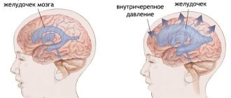
Causes of intracranial pressure
Taking into account the fact that intracranial pressure is formed by the influx of cerebrospinal fluid (CSF) into the ventricles of the brain (special cavities where this substance is located), any violations of this indicator are in one way or another connected precisely with the redistribution of this fluid and nothing else. In turn, there are many reasons contributing to the disruption of the outflow or inflow of cerebrospinal fluid:
- Disturbance of metabolic processes when cerebrospinal fluid is not absorbed into the blood;
- Spasm of the muscular wall of blood vessels, preventing normal vascular circulation;
- An increase in the volume of circulating blood, which leads to an increase in the amount of cerebrospinal fluid;
- Insufficient oxygen supply to the brain;
- Meningitis;
- Hemorrhagic cerebral infarction;
- Inflammatory processes occurring in the central nervous system;
- Neoplasms of various types;
- Intoxication;
- Migraine.
Frontit
Frontal sinusitis (frontal sinusitis) is an inflammatory disease of the frontal paranasal sinus. This type of sinusitis is the most severe. There are forms of acute and chronic frontal sinusitis.
Symptoms of sinusitis
Acute frontal sinusitis, symptoms:
- pain and swelling around the nose and eyes;
- increased pain when tapping in the projection area of the inflamed sinuses;
- heavy breathing due to inflammation of the nasal passages;
- runny nose with thick yellow or green mucus;
- increase in body temperature to 38-39 degrees;
- severe headache (minor relief occurs when lying down);
- pain radiating to the ears and teeth;
- fear of light;
- severe weakness;
- sometimes sore throat, difficulty identifying odors, decreased pungency of taste.
Chronic frontal sinusitis, symptoms:
- aching headache;
- purulent, unpleasant-smelling nasal discharge in the morning;
- slight increase in temperature;
- difficulty breathing through the nose;
- sputum discharge in the morning.
Causes of frontal sinusitis
The following reasons for the development of frontal sinusitis are distinguished:
- viral, bacterial or fungal infection;
- complication after influenza, ARVI, etc.;
- getting foreign objects into the nose;
- long-term infectious or allergic rhinitis (rhinitis);
- deviated nasal septum;
- adenoids;
- allergy;
- nasal polyps.
Treatment of frontal sinusitis
How to treat frontal sinusitis? Definitely under the supervision of an otolaryngologist! The disease is not only difficult for many patients to tolerate, but also has dangerous complications, including orbital abscess, meningitis, sepsis, etc.
Treatment of sinusitis is aimed at eliminating infection in the sinuses and stopping inflammation. Medicines will help relieve swelling, improve ventilation of the sinuses and lead to the discharge of contents from them. If the disease is viral in nature, then antibiotics for frontal sinusitis are mandatory!
The following antibiotics are used to treat sinusitis:
- penicillin antibiotics (semi-synthetic or synthetic amoxicillin preparations);
- cephalosporin antibiotics;
- macrolide antibiotics (they do not affect the intestinal microflora);
- local antibiotics in the form of nasal drops, nasal spray, aerosol;
- homeopathic medicines;
- symptomatic remedies for frontal sinusitis in the form of vasoconstrictor nasal drops, antipyretics and anti-inflammatory drugs.
In case of severe frontal sinusitis and insufficient effectiveness of conservative treatment, sinus lavage using the method of displacement and puncture is prescribed.
1 Rhinoscopy in MedicCity
2 Consultation with an ENT specialist in MedicCity
3 ENT consultation in MedicCity
Prevention of frontal sinusitis
To prevent frontal sinusitis, you need to monitor the state of your immune system, promptly eliminate foci of inflammation in the ENT organs, harden your body, and lead a healthy lifestyle.
Treatment of increased intracranial pressure at ON CLINIC Ryazan
If ICP is a secondary disorder and represents one of the complications of any disease, it is necessary first of all to treat the primary pathology. In other cases, symptomatic treatment of ICP is performed.
There are conservative and surgical treatments for this pathology. Conservative - taking medications prescribed by the attending physician that enhance the outflow of fluid according to the regimen prescribed by the attending physician. These can be either regular diuretics or anti-inflammatory steroids. Physiotherapy and massage have also been proven to treat increased intracranial pressure.
Surgical intervention is performed in the most severe cases of ICP. Usually this is bypass surgery - the installation of a special shunt through which excess fluid moves away from the brain, rushing into the abdominal cavity.
Untreated ICP in a timely manner can lead to such serious complications as stroke, partial and complete paralysis, impaired coordination, reflexes, speech, as well as loss of vision and mental problems. If you experience symptoms that may be signs of a deterioration in the flow of cerebrospinal fluid from the brain, consult a doctor as soon as possible! “ON CLINIC Ryazan” - Your health is our concern!
Ethmoiditis
Ethmoiditis is an acute or chronic inflammation of the mucous membrane of the cells of the ethmoid labyrinth (anatomical labyrinth in the bridge of the nose). Ethmoiditis is bacterial or viral in nature.
There are acute and chronic ethmoiditis. Acute ethmoiditis accompanies influenza, rhinitis and is complemented by inflammation of the paranasal sinuses.
Ethmoiditis in adults affects both the frontal and maxillary cavities. With weak immunity, the acute form of ethmoiditis turns into long-term chronic ethmoiditis with periods of exacerbation and remission.
Polypous ethmoiditis is characterized by the appearance of polyps in the mucous membrane of the ethmoid labyrinth of the forehead. Polypous ethmoiditis may appear after chronic, allergic rhinitis.
Catarrhal ethmoiditis occurs due to the activity of viruses. It is characterized by increased lacrimation, weakness, nausea, dizziness, swelling in the bridge of the nose, and fever.
Ethmoiditis in children is a very serious disease. The infection spreads very quickly due to the anatomical structure of the ethmoid labyrinth. The development of ethmoiditis in children requires urgent hospitalization.
Symptoms of ethmoiditis
Acute catarrhal ethmoiditis. Symptoms
- pain in the bridge of the nose and at the wings of the nose;
- heavy breathing through the nose;
- loss of smell;
- headache, weakness;
- profuse nasal discharge, which gradually becomes purulent;
- temperature rises to 38 degrees;
- In children, the inner corner of the eye socket also swells and turns red.
In the acute form, primary and secondary ethmoiditis are distinguished.
With primary ethmoiditis, anxiety, vomiting, dyspepsia and toxicosis appear, the temperature is 39-40 degrees.
Secondary ethmoiditis is more severe and develops faster. The patient is in extremely serious condition with pronounced septic symptoms. The eyelids become swollen and cyanotic, swelling of the conjunctiva and noticeable protrusion of the eyeball are observed, and nasal breathing becomes difficult.
Chronic ethmoiditis. Symptoms
- headaches that are difficult to register by localization;
- weakness, rapid fatigue of the patient;
- soreness in the bridge of the nose when pressed and painful points at the wings of the nose;
- purulent discharge with a nauseating odor;
- extensive mucus in the nasopharynx, which is difficult to spit out;
- emerging polyps.
Complications after ethmoiditis:
- meningitis;
- encephalitis,
- intraocular and intracranial pressure;
- destruction of the ethmoid bone.
Treatment of ethmoiditis
Treatment of ethmoiditis in acute form is predominantly conservative. It is necessary to ensure the outflow of mucus with the help of vasoconstrictors and physiotherapeutic procedures.
Treatment of ethmoiditis in chronic form is predominantly surgical.
1 MRI in MedicCity
2 Laboratory diagnostics in MedicCity
3 Ultrasound examination of the paranasal sinuses in MedicCity
Diagnostic methods
It is impossible to measure intracranial pressure at home. The only thing a patient can do to help himself is to detect the symptoms in time and consult a doctor for a detailed diagnosis. It is worth understanding that the procedures for measuring ICP are complex and require special equipment and sufficiently qualified medical personnel. All these conditions are available at the Clinical Institute of the Brain, which specializes in the diagnosis and treatment of pathologies of nervous activity.
The only way to accurately determine intracranial pressure is to puncture the cerebrospinal fluid. The technique is invasive and is used only in complex cases. To do this, it is necessary to make a puncture in the lumbar region (into the spinal cord canal) or the ventricles of the brain. The cerebrospinal fluid that is constantly circulating in these spaces will begin to flow out, and its pressure can be measured. The value is measured in mm of water column, and its norm is from 60 to 200 mm. These data are indicative if the patient is in a supine position.
There are also additional diagnostic techniques that allow you to assess the condition of the brain, its ventricles and vascular bed without invasive intervention. These include:
- Ultrasound of the brain - the procedure is performed only for children who have not had fusion of the fontanelle, and in adults it is impossible due to the density of the bones of the skull;
- CT or MRI of the brain - analysis can be done at any age, while the data is quite informative and allows you to obtain a complete three-dimensional image of any area under study;
- echoencephalography is a type of ultrasound examination that can be used to determine the degree of filling and pulsation of the cerebral arteries.
It is impossible to diagnose increased intracranial pressure based on the clinical picture alone. However, all this data must be provided to the doctor during the initial examination. Based on them, specialists from the Clinical Institute of the Brain will prescribe all the necessary diagnostic stages, which will allow a full assessment of the patient’s condition.
Diagnosis of sinusitis using modern techniques
To confirm the diagnosis of sinusitis, the following types of examination are used:
- Video endoscopy of the nasal cavity and nasopharynx to identify features of the anatomical structure and determine predisposing factors for the development of sinusitis;
- radiography of the paranasal sinuses;
- Ultrasound examination of the paranasal sinuses is a safe method with no contraindications, used to diagnose sinusitis and monitor the treatment process;
- CT, MRI - according to indications;
- laboratory diagnostics according to indications in full.
Treatment of sinusitis at MedicCity
Conservative methods of treating sinusitis
If you are concerned about how to treat sinusitis in Moscow, be sure to contact the MedicCity specialists! Our clinic provides treatment for sinusitis without puncture and without pain. However, non-surgical treatment of sinusitis is possible only at the initial stage. Don't waste time!
In the vast majority of cases, treatment of sinusitis in our clinic is carried out without a puncture.
- Using YAMIK (sinus catheter). The YAMIK method is the use of a device called the “YAMIK sinus catheter.” Using the YAMIK sinus catheter, controlled pressure is created in the nasal cavity and the purulent contents of the sinus are pumped out through the natural anastomosis (openings), and then a medicinal substance (antibiotics, mucolytics) is administered.
- Rinsing the nose and paranasal sinuses using the moving method (“cuckoo”). It is carried out using a special suction - an aspirator; in the process, pathological contents are removed from the nasal cavity and sinuses and the drug is injected into the sinuses.
- Inhalation therapy using a special inhaler PARI SINUS. This method is based on the introduction of microparticles of the drug into the affected paranasal sinuses through a pulsating supply of an aerosol. In this case, the aerosol of the medicinal substance is deposited in the sinuses and has an effect directly at the site of inflammation.
All proposed methods for treating sinusitis are painless and effective.
When using combined treatment, complete recovery in case of acute sinusitis is achieved within 7-10 days.
If puncture treatment is necessary, it is possible to install special catheters in the sinus, which eliminate the need for repeated punctures.
Hydrocephalus - symptoms and treatment
To date, the method of surgical treatment of hydrocephalus has proven its effectiveness.
Two types of surgical interventions are used: liquor shunting and neuroendoscopy. Such operations are performed by a neurosurgeon, i.e. a surgeon specializing in the treatment of diseases of the brain, spinal cord and peripheral nervous system.
CSF shunt surgery
In a CSF shunt, a thin tube called a shunt is inserted into a ventricle of the brain. Excess cerebrospinal fluid contained in the brain flows through the shunt to another anatomical region of the human body.
When installing the drainage end of the system into the abdominal cavity, the shunt is called ventriculoperitoneal . In this case, excess cerebrospinal fluid entering the abdominal cavity is absorbed into the bloodstream.
When the draining end of the system is installed into the chamber of the heart (usually the right atrium), the shunt is called ventriculoatrial . They are typically implanted in children because growth has less impact on their functioning and requires replacement less often, unlike ventriculoperitoneal shunts.
Lumboperitoneal are rarely used . They drain cerebrospinal fluid from the subarachnoid space of the lumbar spinal cord into the abdominal cavity.
In the system of tubes through which cerebrospinal fluid flows from the brain, there is a valve with a given throughput. Depending on the cerebrospinal fluid pressure, an appropriate valve is selected for the patient before the operation, which makes it possible to control the outflow of cerebrospinal fluid into the abdominal cavity, i.e. having a given capacity. The neurosurgeon planning surgery determines the pressure of the cerebrospinal fluid in the brain and selects the appropriate valve. It usually appears as a raised “bump” under the scalp.
Today, two types of valves are used:
- with given (preset) parameters that have a certain throughput for cerebrospinal fluid;
- adjustable magnetic valves. When choosing such valves, the attending physician, using special equipment, can remotely, without making additional incisions, change the pressure of the valve and achieve an optimal clinical result, eliminating such adverse side effects as insufficient or excessive drainage of the cerebrospinal fluid.
CSF bypass operations are performed under general anesthesia and take from one to two hours. After surgery, patients usually remain in the hospital for several days.
If the outflow of cerebrospinal fluid through the shunt is disrupted or infection occurs, repeated surgery may be required.
Endoscopic ventriculostomy of the third ventricle
This operation is an alternative to liquor shunt surgery. Instead of installing a shunt, the surgeon creates a hole in the lower wall of the third ventricle and creates a bypass for the outflow of cerebrospinal fluid to the surface of the brain, where its unhindered absorption occurs.
This operation is not universal for all patients with hydrocephalus, but can be used to block the cerebrospinal fluid pathways - occlusive hydrocephalus. In this case, the cerebrospinal fluid flows through an artificially created hole - bypassing the clogged cerebrospinal fluid pathways.
The operation of endoscopic ventriculostomy of the third ventricle is performed under general anesthesia. The neurosurgeon makes a burr hole in the skull with a diameter of about 10 mm and uses an endoscope to examine the ventricles of the brain from the inside. An endoscope is a long, thin tube with a light source and a miniature video camera at the end.
Through the channel located inside the endoscope, it is possible to carry out special surgical instruments to perform operations on the deep structures of the brain. After installing the endoscope into the third ventricle, a hole is formed in its lower wall. Through the newly formed anastomosis, cerebrospinal fluid enters the subarachnoid space. After removing the endoscope, sutures are placed on the aponeurosis and skin. The duration of the operation is about one hour.
The risk of infectious complications is much lower after endoscopic surgery compared to cerebrospinal fluid shunting.
Endoscopic ventriculostomy of the third ventricle has no advantages over cerebrospinal fluid shunting in long-term follow-up. After endoscopic intervention, as well as after cerebrospinal fluid shunting, hydrocephalus can develop again, even several years after the operation.
Treatment of normal pressure hydrocephalus
In case of normal pressure hydrocephalus, which usually develops in older people, the condition can be improved by performing a cerebrospinal fluid shunt operation. Although not all patients with this diagnosis have surgical treatment that is effective.
Due to the risks associated with any surgery, specific tests (stopping lumbar drainage and/or performing a lumbar infusion test) are necessary to assess the potential benefit of surgery, which should outweigh the risk of adverse effects.
According to the literature, more than 80% of patients with normal pressure hydrocephalus who responded positively to preliminary testing reported significant improvement after ventriculoperitoneal shunting. Visible clinical improvement after surgery usually occurs within a few weeks or even months.
A timely and correct diagnosis is the key to successful treatment, even in patients who have suffered from hydrocephalus for several years.[1][7]