Definition
Radiculopathy is a symptom, not an independent disease, a symptom of a disease of the peripheral nervous system, the cause of which in most cases is protrusion or herniation of the intervertebral disc and arthrosis of the vertebral joints. And before continuing, I would like to start with the anatomy of the spine and the structure of the nervous system.
According to the classification of vertebrae, free vertebrae are distinguished:
- cervical vertebrae – 7 pcs.
- thoracic vertebrae – 12 pcs.
- lumbar vertebrae – 5 pcs.
and fused:
- sacral vertebrae – 5 pcs.
- coccygeal vertebrae – 3-5 pcs.
A distinctive feature of the cervical vertebrae is the presence of an opening in the transverse process - the vertebral arteries pass through it. Another distinctive feature is the atlas and axial vertebrae (1st and 2nd vertebrae), they are atypical, they lack a body, spinous and articular processes. 3 - 7 cervical vertebrae - typical.
In any typical vertebra, the body, arch and process are distinguished, and their connections are distinguished according to the parts of the vertebra.
The vertebral bodies are connected to each other through intervertebral discs, anterior and posterior longitudinal ligaments.
The arches are connected by the ligamentum flavum.
Among the processes, spinous, transverse and articular (upper and lower) are distinguished. The spinous processes are connected by the interspinous ligament, the transverse ones by the intertransverse ligament, and the articular processes by the intervertebral joints.
All vertebrae form the spinal column among themselves, and when the vertebrae connect, intervertebral foramina are formed (right and left), through which spinal nerves and vessels pass.
The spinal column does not occupy a strictly vertical position; it has physiological curves. The curve facing backward is called kyphosis, and the curve facing forward is called lordosis.
The spinal cord located inside the spinal canal begins from the foramen magnum and ends to the first lumbar vertebra (LI) in men, 2 lumbar vertebra (LII) in women.
There are 124 roots extending along the spinal cord. 62 posterior and 62 anterior, from which 31 pairs of spinal nerves are formed.
Having understood the anatomy of the spine and the formation of nerve roots, you can move on to the disease.
All back pain can be divided into specific, nonspecific and radicular.
So, radiculopathy is a disease caused by compression (damage) of a nerve root, causing radicular pain in the back. Compression occurs due to the fact that the intervertebral disc, which performs a shock-absorbing function in the spine, begins to protrude under external loads and a non-functioning muscle corset. Thus forming a protrusion, and subsequently a herniation of the intervertebral disc.
For understanding, here are statistics on the prevalence of back pain:
- specific – 85% (due to muscles, ligaments, tendons, small joints),
- nonspecific – 10% (vertebral fracture, tuberculosis, osteomyelitis, abscess, spinal stenosis, neoplasm, ankylosing spondylitis, etc.)
- radicular up to 4%.
In other words, the prevalence of radiculopathy is 4% of all back pain syndromes.
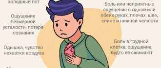
Types of cardialgia
Psychogenic cardialgia progresses in a person against the background of depression or severe emotional shock. The patient exhibits the following symptoms:
- burning and pain in the area of the projection of the heart and in the left hypochondrium. The person notes that he either has a feeling of fullness in his chest, or, conversely, emptiness;
- the pain is constant, throbbing;
— the sensitivity of the skin in the area of the left nipple increases;
- with the psychogenic form of the disease, pain can radiate not only to the neck, spinal column or lower back, but also to the genitals.
Often the pain syndrome is accompanied by the following unpleasant sensations in certain parts of the body: tingling; crawling; numbness; tingling.
Vertebrogenic cardialgia develops when the cervical spine is affected. Pain syndrome manifests itself when the roots of the nerves that exit this part of the spine are compressed. These nerve fibers have a reflex effect on the heart and coronary blood vessels, as a result of which pressing or aching pain occurs in the area of the projection of the heart muscle.
Symptoms of radiculopathy
On the torso, areas of the skin with increased pain sensitivity are divided into certain areas - dermatomes, or they are also called Zakharyin-Ged zones, after the names of the clinical researchers who discovered this phenomenon - Russian therapist G.A. Zakharyin and the English neuropathologist G. Ged.
With radiculopathy, a peripheral version of sensitivity disorder occurs. It is characterized by disorders that occur when sensory pathways are damaged (peripheral nerves, plexuses, roots). And peripheral paralysis or paresis, which is a disorder of voluntary movements. As a result, a sensitivity disorder occurs in the corresponding dermatome. And the muscles innervated by the affected root become weak and hypotonic, which contributes to their atrophy.
Clinical manifestations of radiculopathy are characterized by a sudden onset, with constant or periodic shooting, piercing, intense pain, which at least occasionally radiates to the distal zone of the dermatome, the corresponding affected nerve root.
The pain syndrome can appear and intensify with movement, straining, lifting weights, sitting in a deep chair, staying in one position for a long time, coughing and sneezing and weakens with rest, especially if the patient lies on his healthy side, bending the affected leg at the knee and hip joints. Initially, the pain may be dull and aching, but it gradually increases, and sometimes it can immediately reach its maximum.
During the examination, the patient often takes a forced position. Movement in the affected segment of the spine and the limb innervated by it is sharply limited. On palpation, pronounced tension of the paravertebral muscles is noted.
In differential diagnosis, a neurological examination of the patient is of great importance. Due to damage to the nerve root, its function is impaired, therefore, radiculopathy is characterized by a violation of pain, temperature, vibration and other sensitivity (including in the form of paresthesia, hyper- or hypoalgesia, allodynia, hyperpathia) in the corresponding dermatome, a decrease or loss of tendon reflexes, closing through the corresponding segment of the spinal cord, hypotension and weakness of the muscles innervated by this root.
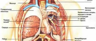
Pleural pain
Pleural pain is a variant of nociceptive somatic pain associated with irritation of the parietal layer of the pleura. As a rule, pleural pain is acute, quite clearly localized and intensifies with deep breathing, coughing or sneezing. The causes of pleural pain include a wide range of diseases of both the pleura itself and adjacent organs (Table 3).
. The main causes of pleural pain.
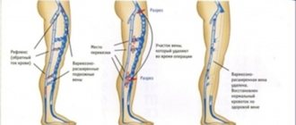
If you suspect the pleural nature of the pain, you should evaluate the presence and severity of accompanying symptoms (shortness of breath, cough, hemoptysis, fever), and when directly examining the patient, pay special attention to the presence of the following signs: • increased venous pressure in the jugular veins; • paradoxical pulse; • displacement of the trachea; • pain on palpation of the chest; • change in percussion sound; • pericardial friction noise; • weakening of breathing; • crepitus or moist fine bubbling rales; • signs of deep vein thrombosis of the legs.
Mandatory laboratory and instrumental studies in the presence of pleural pain are a general blood test, an ECG (sinus tachycardia, decreased amplitude of the QRS complex, changes in the right heart, ST segment elevation or depression of the PQ interval), as well as chest x-ray in two projections ( pneumothorax, infiltrative or focal changes in the lungs, pleural effusion, rib fracture) [24, 25].
Chest pain occurs in up to 50% of patients with pulmonary embolism. The diagnosis of acute massive pulmonary embolism (PE) is usually obvious (in the absence of other diseases and risk factors). At the same time, asymptomatic episodes of pulmonary embolism often go unnoticed in a timely manner. The most typical clinical manifestations are shortness of breath, tachypnea, tachycardia, less often cough and hemoptysis, as well as clinical signs of deep vein thrombosis of the lower extremities. In 80–90% of patients with pulmonary embolism, one or more predisposing factors can be identified, the presence of which helps the clinician in establishing the correct diagnosis. Risk factors also facilitate decision-making in the event of “dubious” research results.
The main risk factors for the development of pulmonary embolism: • immobility of the patient due to various reasons: postoperative period, heart failure, contrast-enhanced tomography. Suspicion of pulmonary embolism is an indication for emergency hospitalization [26].
Pericardial pain can be clinically difficult to distinguish from pleural pain. Direct examination of the patient may reveal increased pain in the supine position and relief in the upright position, as well as a pericardial friction rub. However, information obtained from clinical examination is usually insufficient. In this case, the supporting diagnostic signs are the identified characteristic changes on the ECG (Fig. 2) and echocardiography data [27].
The basis of symptomatic therapy for BS in the chest of a pleural or pericardial nature are NSAIDs.
Pathogenesis
The onset of the development of the disease is two factors that are related to each other: mechanical irritation of the root and/or spinal ganglion and inflammatory changes in the perineural tissue that occur as a result of penetration of the disc into the epidural space.
In this case, factors of root compression can be both disc herniations and bone growths (uncovertebral, spondyloarthritic). Compression can also be caused by hypertrophied ligaments and periarticular tissues, vascular structures (epi- and subdural hematomas, arteriovenous malformations, epidural hemangiomas).
Until now, “blank spots” remain in the pathophysiological concept of radicular pain. It is assumed that the basis of radicular pain is axonal dysfunction caused by various etiological factors, including neural compression, ischemia, damage by inflammatory and other biologically active substances. Spinal roots (unlike peripheral nerves) have a weak blood-neural barrier, making the axon more susceptible to compression injury.
Increased vascular permeability due to mechanical compression of the root leads to endoneurial edema. As a result, a precedent arises that prevents full capillary blood supply and the formation of interneural fibrosis. The spinal root receives up to 58% of its nutrition from the surrounding cerebrospinal fluid (CSF). Perineural fibrosis prevents the complete supply of axonal tissue with nutrients due to diffusion from the CSF, which also contributes to increased sensitivity of the fiber to pressure.
Studies using experimental root compression have shown that even at minimal pressure (5–10 mm Hg) venous blood flow ceases.
The occlusion pressure of the radicular arterioles is significantly higher (approximately corresponds to mean blood pressure), but depends on the potential for venous stasis. Ischemia of nerve fibers or venous congestion leads to biochemical changes that can maintain pain sensations.
Work with experimental root compression demonstrates that compensatory diffusion of nutrients from the CSF is impaired in the setting of epidural inflammation or in the presence of fibrosis.
Recent studies have shown that degenerative changes in the nucleus pulposus and annulus fibrosus can lead to local neural changes and the synthesis of algogenic agents such as metalloproteinases, tumor necrosis factor (TNF), interleukin (IL)-6 and prostaglandin E2. Pathogenetically, the pain syndrome in radiculopathy is of a mixed nature, including nociceptive and neuropathic components.
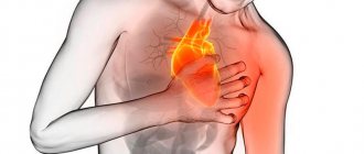
Symptoms of cardialgia
As the disease progresses, the following symptoms appear:
- pain syndrome localized in the left half of the chest, behind the sternum. In rare cases, pain also occurs in the armpit area. It is noteworthy that pain directly depends on the position of the person’s body. For example, it can intensify if a person bends forward or raises his left arm up;
- sleep disturbance; sense of anxiety;
- violation of the swallowing reflex;
- darkening of the eyes;
- the patient cannot take a full breath, so he has a feeling of lack of air;
- in severe cases, the development of pre-fainting or convulsions is possible;
- if the symptoms of the disease appear in a state of complete rest, then this may indicate the progression of neurocircular dystonia. In this case, the following symptoms join the main clinical picture: constant fatigue, weakness, lethargy, decreased performance.
Classification of the disease
Over the years, changes occur in our body, for example, a baby’s skin is soft and elastic, but at 30 years old it is no longer so. The same thing The same thing happens with our spine. Degenerative and dystrophic processes in the spine contribute to the formation of protrusions and hernias, which can and do lead to radiculopathy in the future.
There are discogenic and vertebrogenic forms of the disease. Vertebrogenic radiculopathy is a secondary type of disease in which the spinal cord root is compressed in a kind of tunnel formed by various pathological processes. This may be soft tissue swelling, tumor, osteophytes, disc herniation.
As the degenerative inflammatory process develops, the tunnel narrows, indentation and severe pain appear.
Depending on the location, the following forms of radiculopathy are distinguished:
- cervical;
- lumbosacral;
- mixed.
The disease can occur in adults of any age; if left untreated, the disease can lead to disability. Another name for this disease is radicular syndrome. Complex names have not caught on among the people, so you can often hear that a person suffers from radiculitis. Although this name is not entirely correct.
The most common type is lumbosacral radiculopathy. It affects the vertebrae L5, L4, S1. To understand which vertebrae are involved in the process of inflammation, you need to remember that all parts of the spine are designated by Latin names. The sacral region is Os Sacrum, therefore, the vertebrae are designated by the letter S from 1 to 5. The lumbar region is Pars Lumbalis (L1-L5). Cervical region - Pars Cervicalis (C1-C7). Thoracic spine - Pars Thoracalis (Th1-12).
Having familiarized yourself with this classification, it is easy to understand that Th3 means damage to the third vertebra in the thoracic region, and C2 means damage to the second cervical vertebra. The level of damage is determined using an x-ray.
There is an international classification of diseases - ICD 10. It is generally accepted for coding all medical diagnoses. According to the ICD, radiculotherapy is assigned code M 54.1.
Musculoskeletal pain
In cases of cardialgia of a musculoskeletal nature, the cause of “referred” pain is most often pathology of the facet joints of the cervical spine, myofascial pain syndrome (MPPS) of the scalene, trapezius, infraspinatus muscles or the levator scapulae muscle. Local pain is observed with Tietze syndrome and costochondritis. Radiating pain in the chest may be associated with damage to the thoracic spinal cord, thoracic roots, and intercostal nerves. Unlike the cervical and lumbar regions, the thoracic spine is relatively inactive, which predisposes to a much lesser extent to the development of thoracic compression radiculopathies associated with herniated intervertebral discs or narrowing of the spinal canal due to degenerative changes. The main characteristics that make it possible to distinguish this type of pain from angina pectoris include a subjective description of pain (squeezing or pressing), clear localization of pain in the left half of the chest, lack of connection with physical activity, provocation of pain when moving or changing body position, pain on palpation .
MFPS is a chronic syndrome in which local or segmental pain occurs in various areas of the body. A pathognomonic sign of MFPS are myofascial trigger points, which are areas of local pain in the muscle involved in MFPS, upon palpation of which an area of local compaction is revealed, located along the direction of the muscle fibers. Typically, trigger points range in size from 2 to 5 mm. Mechanical pressure on the trigger point causes not only intense local, but also referred pain, and each of these points is characterized by its own strictly defined zone of referred pain and paresthesia. When, when pressing on a trigger point, the patient involuntarily tries to eliminate the stimulus that caused the pain, this phenomenon is described as a “jumping symptom,” which is a characteristic sign of MFPS. There are active trigger points associated with spontaneous pain, and latent trigger points, in the presence of which spontaneous pain does not occur. Most often, local pain is described by patients as intense and sharp, and referred pain as deep and aching. Active trigger points are often accompanied by decreased strength in the corresponding muscle, increased fatigue, and limited range of motion.
In cases where a neurovascular bundle or nerve is located between two trigger points or between a trigger point and a bone structure, conditions for neurovascular compression are created.
Factors contributing to the formation of MFPS: • acute overstretching of the muscle, observed when performing an “unprepared” movement; • prolonged incorrect body position (antiphysiological postures); • exposure to high or, more often, low temperature; • congenital asymmetry of leg length, pelvic ring, foot abnormalities; • nutritional or metabolic disorders; • concomitant psychological disorders (anxiety, depression, sleep disorders).
Tietze syndrome was first described by Tietze in 1921 and is a relatively rare condition characterized by the presence of nonspecific benign reversible painful swelling in the area of the II (60% of cases) or III costal cartilages. In 80% of cases there is a unilateral lesion limited to one costal cartilage. The pain is usually well localized, but can radiate along the entire anterior surface of the chest wall, as well as into the shoulder girdle and neck. There are no redness, increased temperature and other changes in the skin over the affected area. The pain usually resolves spontaneously after 2–3 weeks, but often persists for several months, and residual swelling can persist for up to several years. The disease usually develops at a young or childhood age. The causes are unknown, but most patients have a history of previous episodes of respiratory infections, severe coughing, heavy physical activity, and poor nutrition. Tietze syndrome is often confused with the much more common costosternal syndrome (“anterior chest wall syndrome,” “costochondritis,” “costosternal chondrodynia”), which is one of the most common causes of chest pain. In contrast to Tietze's syndrome, in costosternal syndrome, palpation in 90% of cases reveals multiple areas of pain: in the left parasternal region, below the left mammary gland, in the projection of the pectoral muscles and sternum. There is no local edema. The cartilages of the II and V ribs are most often affected. When the upper costal cartilages are damaged, pain irradiates to the heart area. The pain usually worsens with chest movement. The disease is more common among women over 40 years of age; its pathogenesis remains unknown. For the purpose of differential diagnosis with coronary insufficiency, in addition to the characteristics of pain, which are usually “atypical” for ischemic heart disease, intercostal nerve blocks with the administration of local anesthetics along the posterior axillary line are also used, bringing pronounced relief to patients.
Damage to the sternoclavicular joints is observed in deforming osteoarthritis, rheumatoid arthritis, ankylosing spondylitis, psoriatic and infectious arthritis. Pain in these conditions is usually local, but in some cases it can be projected onto the anterior surface of the chest and in such cases requires a differential diagnosis with diseases of the lungs and heart. The pain intensifies when lifting the shoulder girdle and palpating the sternoclavicular joint. In some cases, swelling and crepitus are observed in the projection of the affected joint.
Sternoclavicular hyperostosis is a relatively recently described disease, manifested by bilateral chronic painful swelling of the clavicle, sternum and first rib. The cause of this condition is unknown, but a possible connection with psoriatic arthritis is being discussed. The diagnosis is based on identifying characteristic radiological changes - hyperostosis, thickening and increased bone density of the clavicles and sternum, ossification of the cartilaginous part of the first rib and the formation of sternoclavicular synostosis. Less common is acceleration of ESR and hypergammaglobulinemia. The disease has a relapsing course. An increase in bone structures and the spread of the inflammatory process in some cases lead to occlusion of the subclavian vein or the development of superior outlet syndrome.
The treatment of musculoskeletal pain is based on the use of NSAIDs, muscle relaxants, blockades, as well as physical therapy and rehabilitation [31–33].
Complications of radiculopathy
In the absence of a proper approach to the treatment and prevention of this disease, radicular syndrome quickly becomes chronic. As a result, changes such as sudden movement, exposure to cold, or stress can trigger an attack of pain.
Another complication may be a persistent impairment of the motor and sensory function of the affected limb and lead to disability. For example, a hernia in the lumbar spine without timely treatment causes peripheral paresis and parylysis of the lower limb, and disrupts the function of the pelvic organs.
Diagnostics
To establish the level of damage, topical diagnosis is of great importance. The main radicular syndromes are presented in the table: [6]
| Spine | Sensory impairments | Movement disorders | Reflexes |
| C3, C4 | Shoulder girdle | Diaphragm | |
| C5 | Anterior shoulder, deltoid region | Deltoid muscle and partially biceps brachii muscle | Decreased reflex from the biceps brachii muscle |
| C6 | Radial surface of the shoulder and forearm, thumb | Triceps brachii, pronator teres, pectoralis major, often pollicis eminence muscles | Decreased or absent reflex from the biceps brachii muscle |
| C7 | Middle and index fingers | Small muscles of the hand, especially the eminence of the little finger | Decreased or absent reflex from the triceps brachii muscle |
| C8 | Little finger | Quadriceps femoris | Decreased reflex from the triceps brachii muscle |
| L3 | Anterior thigh | Quadriceps femoris, tibialis anterior | Decreased knee reflex |
| L4 | Medial surface of the leg | Extensors of the big toe | |
| L5 | Medial surface of the foot, big toe | Foot flexors | Decreased or lost Achilles reflex |
| S1 | Lateral surface of the foot, little toe |
Radiculopathy requires a CT or MRI scan of the affected level of the spine. In order to assess the level of research, it is necessary to find out the symptoms of the disease, neurological status during admission. If the level of the lesion cannot be determined, electromyography is prescribed, which helps to target the affected root, but does not allow the cause to be determined.
If neuroimaging does not reveal atomic changes, it is necessary to study the cerebrospinal fluid to exclude infectious and inflammatory causes, as well as determine the level of glucose in the blood to exclude diabetes.
Treatment of radiculopathy
A neurologist treats herniated discs. Medical specialists have extensive experience in treating such diseases. You can go through all stages of treatment under the supervision of your attending physician, who will answer all your questions.
In most patients with radicular pain, conservative treatment is effective; however, in 2% of patients there are absolute indications (progression of sensory and motor disorders, cauda equina syndrome) for surgical treatment.
In general, we can say that conservative tactics for managing patients with this disease are favorable in most cases and should be considered as a priority in the absence of a verified compressing substrate.
The first stage of treatment is non-steroidal anti-inflammatory drugs (NSAIDs). They have analgesic and anti-inflammatory effects. Corticosteroid (CS) injections may be considered as an alternative to NSAIDs for the treatment of radicular pain. Perineural injections (translaminar, epidural, transforaminal or selective root block) should be performed only after neuroimaging (MRI) of a clinical topical diagnosis. To influence the neuropathic component of radicular pain, some drugs from the group of anticonvulsants (carbamazepine, gabapeptin, pregabalin, lamotrigine) can be used.
According to studies conducted to evaluate the effectiveness of systemic local analgesic drugs lidocaine, mexiletine, tocainide and flecainide showed good results.
Diagnosis of cardialgia
If pain occurs in the area of the projection of the heart, it is important to immediately visit a medical facility for a full diagnosis of this condition, because such a symptom may indicate both cardialgia and the presence of pathologies of the cardiovascular system. Only a qualified specialist will be able to conduct a competent differential diagnosis and prescribe adequate treatment.
The standard diagnostic plan for cardialgia includes the following examinations: echocardiography; X-ray; EGDS of the stomach; ECG; Ultrasound; MRI of the heart; CT scan of the heart.
Forecast and prevention of the disease
It is necessary to recommend that the patient return to normal daily activities as quickly as possible, since maladaptive pain behavior is the main barrier to recovery. In addition, comorbid depression also negatively affects treatment outcomes. If pain persists for >4–6 weeks, it is advisable to add antidepressants to analgesic therapy. Pathogenetically, it is most justified to use antidepressants that act on both neurotransmitter systems (serotonin and noradrenergic) for the treatment of pain symptoms. Tricyclic antidepressants (TCAs), which block the reuptake of serotonin and norepinephrine, have greater potential than selective antidepressants. TCAs are more successful in treating pain symptoms and leading to more complete remission of depression. A new class of drugs, dual-action antidepressants that block the reuptake of serotonin and norepinephrine, have high analgesic efficacy and a more favorable spectrum of side effects than TCAs. The effectiveness/safety ratio is optimal for dual-acting antidepressants (duloxetine, venlafaxine). During the recovery period, active exercises are recommended to strengthen the muscle corset.