© Author: Sazykina Oksana Yuryevna, cardiologist, especially for SosudInfo.ru (about the authors)
Electrical axis of the heart (EOS) is a term used in cardiology and functional diagnostics, reflecting the electrical processes occurring in the heart.
The direction of the electrical axis of the heart shows the total magnitude of bioelectric changes occurring in the heart muscle with each contraction. The heart is a three-dimensional organ, and in order to calculate the direction of the EOS, cardiologists represent the chest as a coordinate system.
When taking an ECG, each electrode records bioelectrical excitation occurring in a certain area of the myocardium. If you project the electrodes onto a conventional coordinate system, you can also calculate the angle of the electrical axis, which will be located where the electrical processes are strongest.
Correct placement of EOS in children
Babies have a strong axis deviation to the right side, which during the first year of life turns into a vertical plane.
This situation has a physiological explanation: the right side of the heart “overtakes” the left in weight and production of electrical impulses. The transition of the axis to normal is associated with the development of the LV. Children's EOS standards:
- Up to a year - the passage of the axis is between +90 - +170 degrees.
- From one to three years - vertical EOS.
- 6-16 – stabilization of indicators to adult standards.
Lexical minimum
As you know, I really love Latin (see my lessons - times two and three). Therefore, in every article that deals with anatomy, I duplicate each term in Latin, and then, at the end of the article, I provide a list of terms used to make it easier for you to learn and remember them.
In this way I would like to create in my readers a certain vocabulary of the Latin language, which will undoubtedly help them in the study of anatomy, surgery and other sciences. I recommend writing down the terms in a notebook dictionary and signing the translation next to it, which you will find in the text of this article
- Cranium
- Musculi masticatorii
- Encephalon
- Lobus temporalis dexter
- Vertebra cervicalis
- Oesophagus
- Trachea
- Glandula thyroidea
- Clavicula
- Etremitas acromialis
- Sternum
- Incisura jagularis
- Ventriculus
- Dorsum
The answer to the question is that we look at the skull in the frontal plane, and it is sawn, of course, in the sagittal plane.
Why do I use a pencil to diagnose or when I don’t need to look for the alpha angle?
Visual determination of the alpha angle
There is another method, the simplest and most beloved by students, of determining the position of the EOS using a pencil. It is not effective in all cases, but sometimes it simplifies the determination of the cardiac axis, makes it possible to determine whether it is normal or whether there is a displacement. So, with the non-writing part, we apply the pencil to the corner of the cardiogram near the first lead, then in leads I, II, III we find the highest R.
We direct the opposite pointed part of the pencil to the R wave in the lead where it is maximum. If the non-writing part of the pencil is in the upper right corner, and the pointed tip of the writing part is in the lower left, then this position indicates the normal position of the heart axis. If the pencil is located almost horizontally, we can assume a shift of the axis to the left or its horizontal position, and if the pencil takes a position closer to vertical, then the EOS is deviated to the right.
Anatomical planes
Many students confuse this basic thing. There are three main anatomical planes: sagittal, frontal and horizontal.
If you don’t remember geometry at all and have no idea what a plane is, imagine that we are talking about a thin but very sharp metal sheet with which we will saw an anatomical object. After we make the necessary cut, we will leave the metal sheet in the same place.
The Internet is full of pictures like this:
The planes are shown here quite clearly and they fit my analogy, only the conventional “sharp metal sheets” here are also multi-colored. The red “sharp leaf” is the sagittal plane. Blue - frontal plane. Green - horizontal plane.
Let's look at each plane with individual examples
1.Sagittal plane
The name of this plane comes from the Latin word sagitta, which means "arrow". Let's look at the illustration of the brilliant Da Vinci: this is exactly what the skull (cranium) will look like if we cut it into two equal halves in the sagittal plane:
The sagittal plane is also sometimes called the “profile” plane. Excellent expression, much better remembered. We cut the preparation to look at its insides in profile. Here is a tablet with a sagittal section of the head:
The sagittal plane divides the human body into right and left halves.
2. Frontal plane
Just in the last article about the masticatory muscles (musculi masticatorii), we looked at a skull sawn in the frontal plane. Let me remind you of this picture:
Let's look at the whole skull from above:
And now let’s cut it in the frontal plane:
The frontal plane divides the human body into the front and back. It's easy to remember without getting confused: "front" is an English word that means "in front", "front". Imagine, for example, a skull and divide it roughly into the front and back. The plane that will separate them will be the frontal one.
3.Horizontal plane
This plane is often found in diagrams and presentations on topographic anatomy. You can also see CT and MRI images in the horizontal plane. For example, this picture of the brain is taken in a horizontal plane. We clearly see a formidable neoplasm in the right temporal lobe (lobus temporalis dexter):
The horizontal plane is well suited to examine in detail the cavity or layered structure of the limb. This is what makes it so popular among representatives of radiation diagnostics and topographic anatomy.
The fascia of the neck is best viewed in a section in the horizontal plane:
So that you don’t get confused, I decided to highlight several formations that are familiar to you:
- The cervical vertebra (vertebra cervicalis) is highlighted in green (in the shape of the letter “T”);
- The bright yellow ring just above is the esophagus (oesophagus);
- Even higher is the red ring - the trachea;
- In front it is covered by a blue horseshoe - the thyroid gland (glandula thyroidea). From this perspective, it is clear why it is called the thyroid.
The horizontal plane divides the human body into upper and lower parts.
Normal in children
ECG of a newborn
In newborns and infants, there is a pronounced deviation of the EOS to the right on the electrocardiogram; by the age of one year, in most children, the EOS moves to a vertical position. This is explained physiologically: the right parts of the heart are somewhat more dominant than the left ones both in mass and in electrical activity, and changes in the position of the heart can also be observed - rotations around its axes. By the age of two, many children still have a vertical axis, but in 30% it becomes normal.
The transition to the normal position is associated with an increase in the mass of the left ventricle and cardiac rotation, during which the fit of the left ventricle to the chest decreases. In preschool children and schoolchildren, normal EOS prevails; the vertical electrical axis of the heart may be more common, and less often the horizontal electrical axis of the heart. Summarizing the above, the norm in children is considered to be:
- Interpretation of ECG in adults and children, norms in tables and other useful information
- during the newborn period, EOS deviation is from +90 to +170
- 1-3 years - vertical EOS
- school age, adolescence - half of the children have a normal axis position.
What to do if EOS displacement is found on the cardiogram?
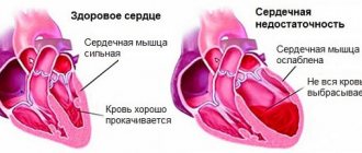
None of the above diagnoses can be made on the basis of EOS displacement alone. The position of the axis serves only as an additional indicator in diagnosing a particular disease. If the deviation of the heart axis is outside the normal range (from 0 to +90 degrees), consultation with a cardiologist and a series of studies are necessary.
And yet, the main reason for the displacement of the EOS is myocardial hypertrophy. The diagnosis of hypertrophy of a particular part of the heart can be made based on ultrasound results. Any disease that leads to a displacement of the heart axis is accompanied by a number of clinical signs and requires additional examination. The situation should be alarming when, with a pre-existing position of the EOS, its sharp deviation on the ECG occurs. In this case, the deviation most likely indicates the occurrence of a blockade.
In itself, the displacement of the electrical axis of the heart does not require treatment; it refers to electrocardiological signs and requires, first of all, to determine the cause of its occurrence. Only a cardiologist can determine the need for treatment.
The concept of EOS norm
The position of the EOS depends on:
- The speed and correctness of impulse movement through the cardiac systems.
- Quality of myocardial contractions.
- Conditions and pathologies of organs that affect the functionality of the heart.
- Heart condition.
For a person who does not suffer from serious diseases, the axis is characteristic:
- Vertical.
- Horizontal.
- Intermediate
- Normal.
The normal position of the EOS is located according to Died at coordinates 0 - +90º. For most people, the vector passes the limit of +30 - +70º and is directed to the left and down.
In an intermediate position, the vector passes within +15 - +60 degrees.
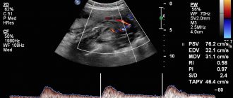
According to the ECG, the specialist sees that the positive waves are longer in the second, aVF and aVL leads.
Vertical plane
Please remember - in anatomy there is no plane with this name!
To avoid confusion, never start listing planes in anatomy with the horizontal one. The word “vertical” may automatically pop out of your mind by association, and then it will seem to your interlocutor/teacher that anatomy has passed you by.
It is best to talk about the horizontal plane at the very end, after the sagittal and frontal.
Let's consolidate knowledge
Let's use another illustration of the magnificent Da Vinci. Try to determine in which plane we are looking at the skull, and in which plane it is sawn. See the answer under the lexical minimum of this article.