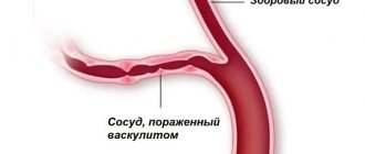
Periarteritis nodosa
- inflammatory damage to the arterial wall of small and medium-sized vessels leading to progressive organ failure with the formation of microaneurysms.
Another common name is polyarteritis nodosa
.
Although vascular diseases were known back in ancient times, conditions with inflammation of blood vessels (vasculitis) as independent clinical forms were described only in the middle of the 19th century.
The first to describe in detail a form of necrotizing systemic vasculitis were Kussmaul and Mayer in 1866, based on the case history of a 27-year-old apprentice tailor. They were the first to use the term “periarteritis nodosa” - periarteritis nodosa. Kussmaul and Mayer considered the condition described as a "unique arterial disease" and distinguished it from infectious disorders, including trichinosis, diphtheria and syphilis.
Even before their report, other authors described aneurysmal dilatations of arteries of inflammatory nature. But in most cases, the information is not enough to describe situations like polyarteritis nodosa. A number of authors believe. that the first case of periarteritis nodosa was described by the Viennese pathologist Karl Rokitansky in 1852. Then, in his article “On some of the most important diseases of the arteries,” he described multiple aneurysmal lesions of the arteries noted at autopsy of a 23-year-old man who was hospitalized with a history of bloody diarrhea and fever with abdominal pain. Rokitansky published macroscopic descriptions of aneurysms in autopsy specimens. He considered the case he described as a type of dissecting aneurysm caused by rupture of the intima and media of small arteries.
In Russia, the first intravital description of polyarteritis nodosa was made in 1928 by Evgeniy Tareev. After further study, chapters on polyarteritis nodosa were published in the monographs “Clinic of Collagen Diseases” (Nesterov A.I., Sigidin Ya.A., 1961) and “Collagenoses” (Tareev E.M., 1965).
Historically, five etiologic hypotheses for polyarteritis nodosa have been studied.
At first, after the description, scientists were most attracted to the hypothesis of the syphilitic origin of vascular lesions, despite the fact that Kussmaul and Mayer clearly rejected the possibility of syphilis in their observations. However, a number of researchers, such as Schmorl, insisted that they simply could not identify the pathogen.
According to the second hypothesis, mechanical factors were assigned key importance in the pathogenesis of periarethritis nodosa: arterial hypertension (Mayer, Klemperer and Ohtani) or congenital weakness of the internal elastic layer of the vascular wall (Eppinger).
According to the third hypothesis, weakening of the vascular wall occurred due to exposure to toxic substances, such as alcohol.
The most popular hypothesis was that arterial damage occurred after an acute infectious disease (streptococcal or staphylococcal, some viral infection) in which the pathogen had a direct or toxin-mediated effect on the vessels. However, for a long time, attempts to detect the pathogen were unsuccessful. Then a new theory appeared.
In 1903, Maurice Arthus described the phenomenon of local anaphylaxis, which later received his name. Until the 1950s it was believed. that the main pathogenetic factor of nodular periartery is precisely the phenomenon of hypersensitivity.
Only in 1947 did the first report of the appearance of polyarteritis nodosa appear in patients who had suffered “serum” hepatitis. By the 1970s, the Gocke and Trepo teams were able to prove the relationship between polyarteritis nodosa and hepatitis B virus infection.
Due to the rarity of the disease, its epidemiology is poorly studied. 0.2-1 cases per 100,000 population are recorded annually. The average age of onset of the disease is 48 years. Men get sick 3-5 times more often than women.
Etiology
There is no clear cause of the disease. Currently, two main factors have been identified:
- drug intolerance;
- persistence of hepatitis B virus.
There are already about a hundred medications that have been associated with the occurrence of periarteritis nodosa.
In 30-40% of patients, hepatitis B surface antigen (HBsAg) or immune complexes including it, as well as other hepatitis B antigens (HBeAg) and antibodies to the HBcAg antigen, formed during viral replication, were detected in the blood. It is known that in France, the incidence of HBV-associated polyarteritis nodosa has decreased from 36% in the early 1980s to less than 5% in the 2000s, due to mass vaccination against hepatitis B.
Also, 5-12% of patients have hepatitis C virus, but its role in causing the disease has not yet been proven. Other viruses are also being considered: HIV, cytomegalovirus, Epstein-Barr virus, rubella, parvovirus B 19, human T-lymphotropic virus type 1, but their role has not yet been proven.
Separate observations have also been published on the occurrence of symptoms of polyarteritis nodosa after administration of a vaccine against hepatitis B and influenza.
Another suspected etiological factor is hereditary predisposition, but it has not yet been possible to establish a connection with a specific HLA antigen.
Manifestations of polyosteoarthrosis
Classic treatment of arthrosis is not always suitable for this diagnosis, since the disease has specific manifestations:
- with generalized arthrosis, three or more joints are affected, most often symmetrical (knee, hip, interphalangeal), Heberden's nodes are noticeable;
- with lesions of the intervertebral joints, pain occurs in different parts of the spine, but secondary symptoms are also possible - dizziness, blurred vision, migraine, indicating compression of the branches of the vertebral artery;
- if polyosteoarthrosis is accompanied by spondylosis of the lumbar and cervical regions with the growth of osteophytes and degenerative changes in cartilage tissue, there is numbness of the limbs, “intermittent” claudication, tinnitus and other unpleasant symptoms;
- sometimes multiple osteoarthritis develops along with periarthritis or tenosynovitis - tissue inflammation, swelling of tendons and capsules.
All this rich symptomatology complicates not only the diagnosis of the disease, but also its treatment.
Polyosteoarthrosis can be accompanied by the most unexpected symptoms
Pathogenesis
Pathogenesis consists of a hyperallergic reaction of the body in response to etiological factors, an autoimmune antigen-antibody reaction (including to the vascular wall), and the formation of immune complexes.
Since endothelial cells have receptors for the Fc fragment of IgG and the first fraction of complement Clq, this facilitates the interaction of immune complexes with the vascular wall. Immune complexes are deposited in the vascular wall, which leads to the development of immune inflammation in it.
The formed immune complexes activate complement, which causes vascular damage, as well as the formation of chemotactic substances that attract neutrophils to the lesion. They phagocytose immune complexes, but in this case they release lysosomal proteolytic enzymes that damage the structures of the vascular wall. Neutrophils are also able to adhere to the endothelium and, in the presence of complement, release activated oxygen radicals, which aggravate vascular damage.
The endothelium also increases the release of factors that promote blood clotting and thrombus formation in the inflamed vessel.
The following are used as agents that alleviate the course of polyosteoarthrosis:
- physiotherapeutic procedures - to relieve swelling, stimulate blood circulation and metabolism, accelerate regeneration;
- manual therapy - to reduce pain;
- acupuncture – to restore mobility;
- other rehabilitation measures (physical therapy, massage, orthopedic treatment, etc.).
Part of the treatment is a proper diet and giving up bad habits.
With a timely approach, even such a complex disease as polyosteoarthritis can be kept under control. The disease begins with pain in the spine and hands, and is manifested by deformation of one of the large joints - the knee or hip. Therefore, you should not ignore any pain symptoms or attribute them day after day to physical fatigue. It is impossible to completely cure arthrosis, and when it comes to multiple lesions, the future is at stake. Be vigilant and attentive, first of all, to yourself - then the disease will not have a chance!
Clinical picture
The disease usually begins with general syndromes: constant fever, progressive weight loss, muscle and joint pain.
Fever occurs in 95-100% of patients, usually of the wrong type, unresponsive to antibiotics, but disappears with corticosteroids. As a rule, it disappears later, when organ pathology appears.
Weight loss with periarteritis nodosa is pathognomic. In some cases, body weight decreases by 30-40 kg in a few months, and the degree of cachexia is higher than in cancer.
Myalgia and, less commonly, arthralgia occur at the onset of the disease. They manifest themselves as characteristic pain in the calf muscles and large joints.
With the development of periarteritis nodosa, five types of organ pathologies are most common. They determine the specific clinical picture of the disease.
Renal vascular lesions occur in 75-90% of patients. The appearance of symptoms indicates a deep stage of the disease. Arterial hypertension usually develops, stable, persistent, leading to severe retinopathy and even loss of vision. Proteinuria (1-3 g per day), microhematuria, and occasionally macrohematuria are observed in the urine.
In some patients, the aneurysmally dilated vessel ruptures with the formation of a perinephric hematoma.
Kidney damage in chronic periarteritis nodosa usually leads to the development of renal failure within 1-3 years.
When the blood vessels of the organs and tissues of the abdominal cavity are affected, symptoms often appear in the early stages of the disease. Abdominal pain is typical, diffuse, persistent, and increasing in intensity. Dyspeptic symptoms are also characteristic: usually diarrhea with a stool frequency of up to 6-10 times a day mixed with blood. Anorexia, nausea, and vomiting occur.
Peritonitis often develops as a result of perforation of an ulcer or intestinal gangrene. Gastrointestinal bleeding is possible.
In 50-70%, damage to the coronary vessels of the heart occurs, although usually not accompanied by anginal pain. Myocardial infarctions develop, mostly small focal ones. Cardiosclerosis progresses rapidly, which leads to rhythm disturbances, conduction disturbances, and heart failure.
Lung damage occurs in about a third of patients with periarteritis nodosa. Manifested by bronchospasms, hypereosinophilia, eosinophilic pulmonary infiltrates.
The development of vascular pneumonia is typical, characterized by a cough with a scanty amount of sputum, occasionally hemoptysis, and increasing signs of respiratory failure.
X-ray examination shows a sharp increase in the vascular pattern, reminiscent of congestive lung, infiltration of lung tissue, mainly in the hilar zones.
Damage to the peripheral nervous system occurs in approximately half of patients. Manifested by the development of asymmetric mono- or polyneuritis. Sharp pain, paresthesia, and sometimes paresis are observed. The lower extremities are mainly affected. Sometimes a picture of polymyeloradiculoneuritis with paresis of the hands and feet develops.
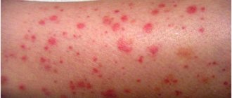
In some patients (15-30%), nodular formations (which give the name of the disease) can be identified along the vascular trunks. Sometimes ulcerative-necrotic changes in the skin occur.
Sometimes damage to peripheral vessels with periarteritis nodosa can lead to soft tissue necrosis and the development of gangrene.
Types and classification of vasculitis
Vasculitis is one of the most serious diseases. The inflammatory process can affect both small vessels of the circulatory system (capillaries, arterioles and venules) and large ones (aorta and its large branches). The affected blood vessels can be located shallowly under the skin, in the internal organs themselves and in any cavities.
Depending on the cause of the disease, there are 2 types of systemic vasculitis:
- primary vasculitis (identified as an independent disease in which the vessels themselves become inflamed);
- secondary vasculitis (appears as a result of other diseases or is the body’s reaction to an infection).
According to the generally accepted classification, primary vasculitis can be divided into three groups, depending on the size of the inflamed vessels:
- inflammation of small vessels (hemorrhagic vasculitis (Henoch-Schönlein purpura), Wegener's granulomatosis (vasculitis), microscopic polyangiitis, cryoglobulinemic vasculitis, Charge-Strauss syndrome);
- inflammation of the middle vessels (periarteritis nodosa, Kawasaki disease);
- inflammation of large vessels (giant cell arteritis (or temporal arteritis, Horton's disease), Takayasu's disease);
There is also isolated “skin vasculitis”, which can be a manifestation of the following diseases:
- (hypersensitive allergic vasculitis, Henoch-Schönlein hemorrhagic vasculitis;
- erythema nodosum, vasculitis nodosum, periarteritis nodosum).
Vasculitis can also affect various human organs:
vasculitis of the brain, vasculitis of the lungs, vasculitis of the lower extremities, vasculitis of the eyes. Eyes with vasculitis can be very damaged (inflammation of the retinal arteries can lead to hemorrhages and partial loss of vision).
Usually people who first encounter this disease are interested in whether vasculitis is contagious. Primary vasculitis is not contagious and is not dangerous to others, but secondary (infectious) vasculitis, which appears against the background of infectious diseases, such as meningitis, can pose an epidemic danger.
Vasculitis during pregnancy can not only provoke delayed fetal development, but also lead to miscarriage.
Diagnostics
The diagnosis is established on the basis of anamnesis (drug allergy, persistence of the hepatitis B virus), a typical polysyndromic clinical picture of the disease, and laboratory test results.
However, laboratory tests for periarteritis nodosa are nonspecific. Their diagnostic value is low; the indicators reflect mainly the degree of activity of the process.
The following diagnostic criteria for periarteritis nodosa are distinguished:
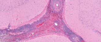
Weight loss of more than 4 kg - a loss of body weight of 4 kg or more since the onset of the disease, not associated with dietary habits Livedo reticularis - a branched change in the skin pattern on the limbs and torso Soreness in the testicles - a feeling of soreness in the testicles, not associated with infection, injury Myalgia , weakness or soreness in the muscles of the legs - diffuse myalgia (excluding the shoulder girdle or lumbar region) or weakness and soreness in the muscles of the lower extremities Mononeuritis or polyneuropathy - development of corresponding neurological manifestations Diastolic blood pressure more than 90 mm Hg. Art. - increased blood pressure Increased level of urea or creatinine in the blood - urea content is more than 14.4 mmol/l (40 mg%) or creatinine is more than 133 µmol/l (1.5 mg%), not associated with dehydration or obstruction of the urinary tract Hepatitis B virus - presence of HBsAg or antibodies to it in the blood serum Arteriographic changes - aneurysms or occlusion of visceral arteries during arteriography, not associated with atherosclerosis, fibromuscular dysplasia and other non-inflammatory diseases Biopsy of small and medium-sized arteries - granulocytic and mononuclear cell infiltration of the vessel wall during morphological examination Presence 3 and more than any criteria allows you to make a diagnosis of periarteritis nodosa.
The reasons for the development of polyosteoarthrosis are:
- hereditary changes in cartilage tissue, which loses elasticity, strength and ceases to withstand dynamic loads;
- weakness of the ligamentous-muscular system;
- diabetes;
- metabolic disorder;
- diseases of the thyroid, parathyroid and other endocrine glands.
Provoking factors are old age (polyosteoarthrosis develops, usually after 50 years), excessive stress on the joints, especially with a sedentary lifestyle. Bad habits and excess body weight also place a person at risk. Injuries can also accelerate the deformation of cartilage, but in this case they are not the main, but rather a provoking factor.
Polyosteoarthrosis of the fingers is passed on from generation to generation in some families.
Treatment
The basis of treatment is glucocorticoids, which are most effective in the early stages of the disease. With long-term use of prednisolone, stabilization of hypertension, progression of retinopathy and renal failure are observed. In acute cases, a paradoxical effect of corticosteroids with the development of multiple infarctions is possible. Also, corticosteroids can dramatically worsen the course of malignant hypertension syndromes. To reduce complications, it is also recommended to use cytotoxic drugs - cyclophosphamide and azathioprine.
Without treatment, the prognosis is extremely unfavorable. The disease occurs at lightning speed, or with periodic exacerbations against the background of steady progression. The cause of death is renal failure, gastrointestinal lesions (especially intestinal infarction with perforation), and cardiovascular pathologies. Often, damage to the kidneys, heart and central nervous system is aggravated by persistent arterial hypertension, which is associated with late complications.
The five-year survival rate without treatment does not exceed 13%; with treatment with corticosteroids it reaches 40%. The labor prognosis is questionable due to the persistence of complications of the disease - peripheral and central paralysis, severe hypertension, heart damage and others.
Typical signs of multiple osteoarthritis
Regardless of the location of the affected joint, the disease makes itself felt with the following symptoms:
- stiffness in movements;
- difficulty moving after sleep (passes within half an hour);
- pain in the affected joints - first after physical activity, and then at rest;
- changing the shape of the joints.
In rare cases, the patient does not even think about treating osteoarthritis, since the symptoms are very smooth or completely absent. However, over time, the disease, alas, progresses.
Is any joint disease arthrosis? Is it possible to cure osteoarthritis quickly? Are chondroprotectors effective in the treatment of polyosteoarthrosis? Am I cured of arthrosis if the pain goes away? A famous rheumatologist answers these and other questions: