Among diagnostic cardiac procedures that are informative, it is worth highlighting echocardiography or ultrasound examination of the heart. This is a hardware procedure performed using high-frequency sound waves. It is indispensable for accurately identifying problems that are directly related to the work of the myocardium and the entire cardiovascular system.
The effectiveness of echocardiography is directly related to the evaluation of its results. Competent interpretation of cardiac ultrasound is a direct path to a speedy diagnosis and drawing up an effective treatment plan. Entrust the solution to the problem to an experienced specialist, contact us for qualified medical support!
What is sudden cardiac arrest?
When the heart suddenly stops, it stops pumping blood.
Sudden cardiac arrest is not the same as a heart attack. A heart attack occurs when one or more of the arteries supplying blood to your heart becomes clogged or blocked. As a result, the heart muscle may suffer. This can be thought of as a problem with the “heart piping”. Sudden cardiac arrest can occur when the heart begins to beat at a dangerously fast rate. This can be thought of as an "electrical" heart problem. Even if the problem with the cardiac pipeline is corrected by angioplasty, bypass surgery, or some other method, the risk of sudden cardiac arrest still remains.
Causes of sudden cardiac arrest.
If you have heart failure or have had a heart attack, there is a chance that your heart muscles are damaged. In this case, the conduction (electrical) system of the heart may also suffer, creating a risk of sudden cardiac arrest. If this situation applies to you, consult with your doctor about the advisability of using an implantable defibrillator for safety reasons.
Sudden cardiac arrest has no warning signs.
Some people may experience palpitations or dizziness, which is a warning sign that a dangerous rhythm disorder may be occurring. But in the vast majority of cases, sudden cardiac arrest occurs without warning. There is no drug that is 100% effective in preventing sudden cardiac arrest. The most effective treatment for sudden cardiac arrest is defibrillation. Defibrillation is the application of a high-energy electrical shock to the heart to restore normal heart rhythm. To prevent death, defibrillation must be performed within a few minutes.
An implantable defibrillator is always ready.
Ambulance crews use external defibrillators. A small device implanted under your skin, an implantable cardioverter defibrillator, can also restore your heart's rhythm and save your life. An implantable defibrillator is always ready to help, monitoring your heart function around the clock. If your implantable defibrillator detects an abnormal rhythm, it automatically provides therapy. An implantable defibrillator is always ready to help you. You can consider it an ambulance that is always with you.
Norms of cardiac echocardiography in children
In pediatrics, standard indicators are directly related to the patient’s body area. It can be determined using a formula using the child’s height and weight data. During diagnostics, review and note information on:
- cavities of the right/left ventricle, as well as the left atrium;
- wall density;
- aorta, especially its ascending section;
- hemodynamics;
- the septum between the heart ventricles.
Competent interpretation of cardiac echocardiography in children requires care and responsibility in all situations.
| Parameters for diagnosing myocardium in newborns | Male | Female |
| Left ventricular EDR | From 19 to 25 mm | From 18 to 24 mm |
| Left atrium diameter | From 13 to 18 mm | From 12 to 17 mm |
| Left ventricle diameter | From 6 to 14 mm | From 5 to 13 mm |
| Left ventricular ESR | From 12 to 17 mm | |
| MZhP (thickness) | From 3 to 6 mm | |
| Right ventricular wall thickness | From 2 to 3 mm | |
| Blood flow speed | About 1.3 m per second | |
| Ejection fraction varies in infants | From 65 to 75% | |
Please note that sometimes after cardiac echocardiography has been performed, interpretation is carried out directly in the diagnostician’s office. The fact is that often the hardware examination is carried out by the attending physician himself, which significantly saves time for the patient and helps the doctor quickly make the correct diagnosis.
Sometimes the list of parameters in the response is different. This happens if the research was carried out with devices of different manufacture and power, that is, devices with different technical characteristics and capabilities.
Do I need an implantable defibrillator?
Doctors of several specialties deal with heart problems. A cardiologist treats problems such as high blood pressure, high cholesterol, and clogged arteries. An arrhythmia specialist focuses on problems with the heart's electrical system that affect how your heart beats. This specialist can determine whether you need an implantable defibrillator.
Typically, candidates for implantable defibrillators have one or more of the following risk factors:
- Heart failure;
- (a heart condition in which the heart's ability to pump blood is reduced);
- Previous myocardial infarction;
- One of the family members had sudden cardiac arrest;
- Low ejection fraction.
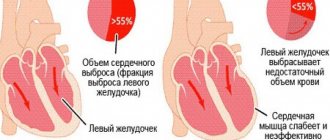
What is ejection fraction?
Ejection fraction is a measure of the percentage of blood that is ejected from the heart into the aorta during each contraction. Typically, at rest, a healthy heart, when contracting, throws out 50-70% of the blood in it. Many people with heart failure have an ejection fraction of less than 40%. A lower than normal ejection fraction means that your heart is not pumping efficiently and cannot meet your body's need for an efficient blood supply.
Why is it important to know your ejection fraction?
Your ejection fraction is one measure your doctor can use to recognize heart failure and assess your risk of sudden cardiac arrest. The latest medical research shows that people who have not had or have had sudden cardiac arrest with an ejection fraction of less than 40% are at greater risk of dangerously accelerated heart rates and sudden cardiac arrest.
How is ejection fraction measured?
Typically, a simple and painless method called echocardiography, or “cardiac ultrasound,” is used to determine ejection fraction. This method, which uses ultrasound to evaluate cardiac function, is widespread and can be performed in most medical institutions.
Interpretation of ultrasound of the heart
In order for the interpretation of cardiac echocardiography to be carried out without errors and completely, the patient’s age, general state of health, and the presence of chronic diseases (pancreatitis, tonsillitis, asthma, vasculitis, etc.) are taken into account. It is impossible to determine the pathology on your own (without knowledge and experience). Only a doctor can correctly assess the answer. There is no place for experiments and guesswork. It is inappropriate to look for answers on the Internet on the websites of companies with a dubious reputation. Trust your health to professionals who provide guarantees and value each patient.
Study parameters
Thanks to ultrasonic waves, you can accurately determine:
- myocardial parameters (sizes of all its parts);
- tissue structure, wall density;
- rhythms, abbreviations, etc.
Imaging will help detect scarring, thrombosis, benign and malignant tumors. The test informs about the condition of the mitral valve, blood volumes and the level of vascular blockage.
By interpreting cardiac echocardiography, the presence/absence of the following diseases can be determined:
- ischemia, when there is a persistent disturbance of blood supply against the background of vascular blockages;
- necrosis, during which tissue death occurs (infarction);
- blood pressure above or below normal (hypo-, hypertension);
- defect, that is, a structural defect of an acquired/congenital type;
- decompensation, when a whole syndrome of failures is noticeable;
- valvular dysfunction;
- rhythmic disruptions;
- rheumatism, when inflammation is observed;
- pericarditis, in which there is inflammation of the membrane;
- stenosis, which indicates a narrowing of the aortic lumen;
- vegetative-vascular dystonia.
Competent image decoding is the basis for success. After all, it is thanks to him that one can establish the fact whether there is a disease or not.
Among the research parameters, several main areas are distinguished. When interpreting cardiac ultrasound in children and persons over 13 years of age, the diameter of the left ventricle and left ventricle, the thickness of the posterior wall of the left ventricle, and more are determined. Each number has a meaning individually. Also, all indicators are taken into account together, because many of them are related to each other and often form a single clinical picture.
How it happens
A painless examination is carried out in a hospital setting or at home, if the situation requires it. The manipulation takes from 20 to 45 minutes. This is a safe way to assess the functioning of the heart muscle and blood vessels, which has no contraindications and is not harmful to health. Step by step it usually looks like this:
- The visitor bares his torso, undresses to the waist and takes a horizontal position, lying on his back with his head towards the diagnostician.
- A special gel is applied to the chest area to help conduct ultrasound waves better.
- A special sensor is used, thanks to which the diagnostician carries out an inspection. The detector is moved slowly across the area being examined to ensure that nothing is missed. The attentiveness of the diagnostician will help with echocardiography decoding.
- The specialist can correct the patient’s position and condition, for example, asking him to hold his breath, roll over, raise his arm, bend his knees, and more.
The response is assessed by a competent specialist, a doctor. It is important that all indicators are taken into account, as well as the individual parameters of the patient’s body. The time of the diagnostic procedure and the state of health, that is, the well-being of the person being diagnosed, are taken into account. Standards and actual figures are reviewed. A comparison is being made. Only after a thorough study can a diagnosis be made and a course of therapy formed. A person who has nothing to do with modern cardiology will not be able to understand the indicators, even if he uses a plate with standard figures for this. Only a properly qualified doctor should be involved in forming a conclusion!
Differences in men, women and children
Normally, data by age and gender is different. EchoCG interpretation helps to analyze the full cycle of myocardial activity (1 systole + 1 diastole). With a heartbeat of 70 beats in 60 seconds, 1 cycle is normally 0.85 seconds.
Adult women and men, as well as children, have different normal values. For example, the heart mass of a representative of the stronger sex aged 25-30 years is about 135 g. In a woman - up to 100 g. The EDV of the left ventricle in men normally reaches 193 ml, in women it does not exceed 136 ml.
The LV wall mass index is an indicator of the ratio of the weight of the organ to the surface area of the human body. The strong half of humanity is characterized by indicators from 71 to 95 g/m2, the fair sex is characterized by a range from 71 to 90 g/m2.
What is an implantable defibrillator?
An implantable defibrillator is a device similar to a pacemaker (artificial pacemaker) that has been used since the early 90s. It is small in size - no larger than a pager - and is implanted under the skin of the upper chest. The device contains a battery and a microcomputer necessary to correct your rhythm. An implantable defibrillator is connected to the heart by thin, insulated wires called electrodes. If the device detects that your heart rhythm is abnormal, it sends electrical signals to restore it. Most implantable defibrillators last 5-7 years before needing to be replaced.
Deviations from the norm
There are a huge number of indicators indicating deviations from the norm. Eg:
- Stenosis is said to occur when the valve opening is reduced in diameter, making it difficult to pump blood.
- If the valve leaflets interfere with the reverse blood flow, perform their natural function inadequately, insufficiency is suspected.
- Pericarditis is detected when, with a normal fluid level of 20-30 ml, all 500 ml are traced.
The list of deviations from the norm and possible diagnoses associated with this can be endless. But you shouldn’t take deciphering an ultrasound of the heart so lightly. Here it is inappropriate to guess and assume without the help of a doctor. Mistakes lead to irreparable consequences. Therefore, experiments should be abandoned.
How does an implantable defibrillator work?
Depending on the programmed mode, the implantable defibrillator may initially send low-energy, painless electrical signals to restore rhythm. If they do not work, a stronger shock is sent. People describe this discharge as a sudden feeling of discomfort, sometimes even painful, but quickly passing. Often everything ends before you understand what actually happened. Another positive thing is that an implantable defibrillator protects you constantly, 24 hours a day.
What happens during the implantation procedure?
Typically, the implantation procedure is performed quickly, safely, and usually under local anesthesia. It does not require open heart surgery, and most patients return home within 24 hours. The following section is a general description of what happens during the implantation procedure. Your case may be different, you can discuss all the details with your doctor.
During implantation.
Typically, patients are sedated before the procedure and do not feel any pain. Your doctor will make a small incision in your upper chest and place electrodes through a vein into your heart. The doctor will then connect the electrodes to the implantable defibrillator and program the device. Next, an implantable defibrillator will be placed under the skin and the incision will be sutured. The doctor will check to see if the implantable defibrillator is working properly.
After the procedure.
After implantation of the device, the patient usually spends the night in the ward and can return home in the morning. You may feel a small bump under the skin where the device is located; this area may be sensitive. It is usually recommended to limit movement of the arm on the implanted side for 2-6 weeks. You can obtain more detailed care instructions from your doctor. However, you can expect a quick return to your previous activities after surgery. If you have any questions, please ask your doctor or nurse.
Starting life with an implantable defibrillator: the first six weeks.
It's time to start living a full life again. We hope it will be filled with hope and confidence. Of course, until healing occurs, you may experience some discomfort. Also, in order not to cause displacement of the electrodes, you may have to slightly limit the movements of your left hand. Typically these restrictions are recommended for 2-6 weeks. Your doctor will tell you in more detail what movements and strains you should avoid. If you have questions about what you can or cannot do before you recover, don't hesitate to ask your doctor or nurse. Your family and friends will be interested in hearing about your implantable defibrillator. You may want to show the implantation site and talk about the operation. Tell everyone how glad you are to feel recovered from your illness. Now is the time to tell your friends and family about what you are planning to do soon. Choose one of your favorite activities and make plans, think about how you will do it again. Believing in yourself and doing enjoyable, enjoyable things makes a big contribution to the healing process.
Living with Success: Achieving the Confidence You Need.
A new device for your heart will help you feel calm and confident. It is always on alert, constantly taking care of your heart 24 hours a day. If your implantable defibrillator detects a rhythm disorder, it sends an electrical impulse to restore it. You can imagine that this is an ambulance that is always nearby. And very soon you will feel the contribution of these positive effects in your everyday life. Remember that you will still need to continue taking your medications and have your device checked periodically by your cardiologist and electrophysiologist (heart rhythm specialist). Either way, your life is in your hands, so enjoy it. Keep making plans. Tell everyone how good you feel, what you have achieved thanks to this, and what you are going to do. For a high quality of life, identify what you enjoy doing and return to an active lifestyle. Always remember that if you have questions about any special activities, such as driving or returning to work, you should consult your doctor.
What happens after the shock: Have a plan.
If your implantable defibrillator detects an abnormal rhythm in your heart, it will automatically deliver therapy—a high-energy shock. Patients who have undergone such therapy describe this discharge as a sudden feeling of discomfort, sometimes even painful, but quickly passing. It also means that the implantable defibrillator has done its job and may have saved your life. Your doctor will give you specific instructions about what to do after the shock. Together with them, develop an action plan that suits you. This plan may include what you will immediately need to do and some of your favorite activities that you can continue to do after the discharge. Keep the plan in a convenient place so you can refer to it. Also, be sure to tell your loved ones and those who care about you about this plan so that they can understand how to help you. It is important to understand that what you were doing during the discharge did not cause it. Typically, you will be able to continue enjoying your favorite activities. Talk with your doctor about when you can return to your activities and whether any changes need to be made. Although the discharge may make you feel temporarily uneasy, it is important to return to your daily activities and focus on the things you love.
You create your own life.
Life is made up of moments, so the more pleasure we get from them, the more fruitful our life becomes. An implantable defibrillator can help you enjoy life more so you can look forward to the future that means so much to you. These can be simple, “earthly” joys, such as walks in the park, working in the garden, or communicating with your loved ones. The important thing is that these moments often make life richer, so hurry up to enjoy them.
Interpretation of EchoCG results in adults
The diagnostic conclusion is made only by the attending physician. The document in written (sometimes electronic) form is transferred to the patient in the diagnostic room, after which it is recorded in the hospital record. The individual indicators of the person undergoing the examination are entered into a special table, which already contains the normal figures for a specific age and gender.
Trying to decipher an ultrasound of the heart on your own is stupid and unsafe. The combination of several deviations may indicate different pathologies. You can’t do without experience and knowledge here.
| Cardiac parameter | Men's answers | Women's answers |
| LVMM—left ventricular myocardial mass | 135-180 g | 95-142 g |
| LVMI—left ventricular mass index | 71-92 g/m2 | 71-88 g/m2 |
| ESD – LV end-systolic size | 31-42 mm | 31-42 mm |
| ESD – LV end-systolic size | 31-42 mm | 31-42 mm |
| EDV - end diastolic size | 46-58 mm | 45-58 mm |
| Volume of fluid in the pericardial cavity | 10-30 ml | 10-30 ml |
| LV wall thickness in diastole | up to 11 mm | up to 10 mm |
| Blood ejection volume during LV systole | 60-100 ml | 60-100 ml |
| Wall thickness of the RV - right ventricle | 5 mm | 4.8-5 mm |
| LA size - left atrium | 18.5-33 mm | 17.5-33 mm |
| LA end-diastolic volume | 50-82 ml | 38-57 ml |
| End-diastolic volume of the RA – right atrium | 20-100 ml | 20-100 ml |
| Thickness of the IVS - interventricular septum in systole | 5-9.5 mm | 5-9.0 mm |
| IVS thickness in diastole | 7.5-11 mm | 7.5-11 mm |
| Aortic opening area | 20-35 mm2 | 20-35 mm2 |
| Thickness of the outer membrane of the pericardium | 1.2-1.7 mm | 1.2-1.7 mm |
Only a cardiologist can evaluate an ultrasound of the heart; the results are interpreted according to standard values, taking into account the general health of the patient, the medications he is taking and other factors. The size, weight, volume and location of the organ, the condition of the valves and tissues are studied. Parameters for contractions, blood vessels and blood flow, wall thickness and other important nuances are recorded. The speed of blood distribution through the chambers of the heart helps to determine Doppler measurements. It is often performed in conjunction with traditional ultrasound examination.