Etiology of pathology
Dyshormonal cardiomyopathy appears due to disturbances in the functioning of the endocrine glands that produce biologically active substances (BAR).
The latter perform a vital role in the body - they regulate metabolism. Therefore, any imbalance in hormone levels leads to organ dysfunction. Involvement of the heart in this process is called myocardial dystrophy. An imbalance in hormonal levels can develop due to:
- neoplasms in the prostate gland;
- pathologies of the thyroid gland (TG);
- menopause;
- decreased testosterone production in men over 50 years of age;
- pathologies of the testicles and ovaries;
- adrenal diseases.
In addition, in some cases, DCM can be diagnosed as an independent disease.
Clinical manifestations
Symptoms of dyshormonal cardiomyopathy may appear gradually or acutely. Subjectively, patients always feel the presence of a serious illness, which does not correspond to reality during the examination.
The main symptom of DCM is cardialgia. The pain is localized at the apex of the heart, occurs suddenly, and can last for a long time. Taking painkillers allows you to relieve pain for a short time, with subsequent return. Patients also note that the discomfort intensifies at night, is not associated with physical activity and can appear at rest.
In addition, such patients complain of:
increased sweating;- shortness of breath;
- frequent surges in blood pressure;
- memory problems;
- dizziness.
With cardiomyopathy of thyrotoxic origin (develops with an excess of thyroid hormones), patient complaints will be different. This form of the disease is characterized by:
- tachycardia (high heart rate);
- inability to concentrate;
- insomnia (insomnia);
- headache;
- dry mouth.
If the prostate gland is involved in the process, men will complain of decreased potency and libido. Due to hyperplasia of the latter, problems with urination appear, and sometimes oliguria (decreased daily urine volume) can be observed.
Cardiomyopathies in children
T.A. RUZHENTSOVA
, Candidate of Medical Sciences,
Central Research Institute of Epidemiology of Rospotrebnadzor, Moscow The article summarizes data on the modern classification, causes and methods of diagnosing cardiomyopathies in children.
Various options for clinical symptoms, electrocardiographic, echocardiographic and biochemical changes are considered. The significance of the results of additional examination methods for cardiomyopathy is assessed. Introduction
Currently, non-coronary heart pathology remains one of the most difficult problems in pediatrics and cardiology. With the development of high-tech diagnostic and treatment methods over the past century, significant improvements in prognosis and an increase in life expectancy in patients with coronary heart and brain disease have been noted. However, morbidity and mortality from myocarditis and cardiomyopathies remained at the same level [1]. According to pathological studies, only about a quarter of cases are diagnosed during life [2]. Among those who died from sudden death syndrome between the ages of birth and 18 years, non-coronarogenic changes in the myocardium are found with a frequency of 20–40% [2–6]. A significant obstacle to the timely formulation of a correct diagnosis is the widespread use both in practice and in the literature of terms that are not included in the International Classification of Diseases, X Revision (ICD X), such as “cardiopathy” and “myocardial dystrophy.” The lack of unambiguous generally accepted diagnostic algorithms and management schemes for this category of patients leads to progression of the disease without adequate treatment and, in some patients, to death.
Terminology issues
The term “cardiomyopathy” was first used in 1957 to designate myocardial diseases of unknown etiology, accompanied by heart failure, cardiomegaly, progressive course and fatal outcome [7]. Later, combined cardiac lesions involving the pericardium and/or endocardium were also included in this pathology. In 1961, Goodwin proposed to distinguish acute, subacute and chronic course. In 1980, a WHO expert committee approved a classification dividing cardiomyopathies into dilated, hypertrophic and restrictive. To denote secondary myocardial changes not associated with damage to the coronary arteries, the term “myocardial dystrophy” has become widespread in the domestic literature. However, with the accumulation of results from high-tech methods for diagnosing myocardial diseases over the past decades, ideas about these nosologies have undergone significant changes. In many cases previously considered idiopathic, according to endomyocardial biopsy, the infectious, autoimmune or dysmetabolic nature of the changes was established [7]. Viral agents and inflammatory infiltration are found in most patients with dilated cardiomyopathy and in some patients with other forms. Dystrophic changes in the myocardium, determined by a decrease in the activity of succinate dehydrogenase, a decrease in glycogen, destruction of myofibrils and mitochondria, often accompany typical leukocyte infiltration in neighboring areas. Sometimes the typical signs of dystrophy after a few days or weeks are replaced by inflammatory ones [8–10]. These data made it possible to identify infectious cardiomyopathies as a separate group. The discovered other changes in cardiomyocytes with corresponding nosologies dictated the need to add arrhythmogenic, peripartum (postpartum), metabolic, autoimmune, and toxic cardiomyopathies to the classification. Thus, today, according to the current ICD X, the following types of cardiomyopathy are distinguished [7, 11]:
I. Idiopathic (dilated - I42.0, obstructive hypertrophic - I42.1, non-obstructive hypertrophic - I42.2, restrictive - I42.5). II. Specific (infectious - I43.0, metabolic - I43.1, with nutritional disorders - I43.2, alcohol - I42.6, with the influence of drugs and other external factors - I42.7, with other diseases - I43.8). Cardiomyopathies include endomyocardial (eosinophilic, Loeffler's endocarditis, endomyocardial, or tropical fibrosis) disease (I42.3) and endomyocardial fibroelastosis (I42.4). Postpartum – O90.3 and cardiomyopathy during pregnancy – O99.4 are included in a separate category.
Idiopathic processes in the myocardium are most often caused by inherited genetic abnormalities or spontaneous mutations leading to disruption of the structure and function of myofibrils.
Today, many specialists dealing with the problems of non-coronary heart diseases have recognized the fact of the staged nature of infectious and inflammatory changes with the transformation of myocardial dystrophy into acute myocarditis, and then, in an unfavorable course, into dilated cardiomyopathy [12–16]. It is not always possible to determine a clear boundary between different types of cardiomyopathies, since there are signs of two or more influencing factors. With specific processes, the ultrasound picture often corresponds to dilated, hypertrophic or restrictive cardiomyopathy. One type can change into another over time. In many cases, it is almost impossible to draw a clear line between myocarditis and cardiomyopathy.
Due to the complexity of diagnosing non-coronary myocardial diseases not only in Russia, but also in other developed countries of Europe and America, the non-specificity of symptoms and the current lack of generally accepted diagnostic criteria, the WHO expert committee in 1997 proposed to use the term inflammatory cardiomyopathy in practice - “ inflammatory cardiomyopathy" [11, 17]. They are recommended to designate all cases of infectious myocardial lesions, their consequences, all acute and chronic myocarditis, including autoimmune, all cases of dilated cardiomyopathy [11, 14, 17, 18]. Inflammatory cardiomyopathy, depending on the predominant pathogenesis, has been proposed to be divided into three forms: infectious, autoimmune and idiopathic. In chronic processes, which, in accordance with WHO recommendations, should be classified as secondary cardiomyopathies, replacement and interstitial fibrosis, hypertrophy of muscle fibers with areas of destruction and focal mononuclear infiltrates are revealed.
Clinical picture of the disease in children
Cardiomyopathy can manifest at any age. All manifestations can be expressed to varying degrees. Some children feel quite well, without making any complaints and without causing concern to their parents [7]. Common symptoms may include lethargy, weakness, fatigue, sleep disturbances, lack of appetite, sweating, dizziness, headaches and muscle pain. Parents often note excessive capriciousness of the child, and sometimes significant disturbances in behavior and perception [19, 20]. Insufficient blood supply to the brain, which develops as a result of a low ejection fraction, may be accompanied by convulsions and fainting. There is isolated evidence that with a long course of myocarditis or cardiomyopathy in children, delayed psychomotor development is noted [13].
Patients may complain of shortness of breath during exercise, and sometimes at rest, palpitations. Associated rhythm disturbances, which can give sensations of interruptions and pauses in the work of the heart, episodes of tachycardia or tachyarrhythmia lead to an increase in symptoms of heart failure [13, 21]. The pain has a different character: stabbing, aching, and when there is insufficient blood supply to the myocardium, pressing. In some patients, the pain syndrome may be mild and feel like discomfort. Damage to the posterior diaphragmatic parts of the left ventricle can cause pain in the upper abdomen, nausea, and vomiting, which often represents a rather difficult differential diagnostic task, especially when abdominal and cardiac pathologies are combined. A decrease in blood supply to the abdominal organs due to a decrease in the ejection fraction of the left ventricle leads to an increase in stool up to 5 times, and in children under 1 year of age up to 10 times a day [13, 19]. The stool, as a rule, becomes unformed; in severe myocarditis or cardiomyopathy, streaks of blood may appear. The pain syndrome can spread to all parts of the abdomen. With prolonged course, constipation may occur as a result of the development of ischemic colitis. An increase in pressure in the vascular bed of the pulmonary circulation against the background of a decrease in the pumping function of the left parts of the heart leads to the appearance of a cough in these children, which can be either dry or wet with the discharge of a small amount of whitish sputum [13]. The severity of manifestations of heart failure, as a rule, determines the prognosis in such children.
When examining the patient, changes in the skin characteristic of heart failure are revealed: pallor, acrocyanosis, marbling. In severe cases, there may be swelling of the legs. If the child is in a horizontal position most of the time, then the swelling spreads to the lumbar region, the anterior abdominal wall, arms and face [22, 23]. When palpating the area of the cardiac impulse, its shift to the left and weakening are noted, which indicates an expansion of the cavities of the heart. Percussion is determined by an increase in absolute dullness, mainly to the right, due to the right ventricle, which is more often involved in the infectious process in children, and/or an expansion of the boundaries of relative dullness to the left by 1–2 cm due to hypertrophy or dilatation of the left ventricle. An upward increase in the boundaries indicates involvement of the left atrium in the process. In some cases, the right border of the relative dullness of the heart is determined in the form of a steep arc with a shift to the right, which indicates changes in the right atrium. Dystrophic and hypoxic damage to the myocardium can, like inflammatory ones, lead to a decrease in ventricular contractility and expansion of the chambers of the heart. With adequate therapy, these patients experience rapid normalization of size. However, the lack of timely correction of disorders can lead to the destruction of cardiomyocytes and the formation of irreversible foci of cardiosclerosis. Auscultatory symptoms may vary. In the initial period of the infectious-inflammatory process in the myocardium, pronounced loudness and clarity of tones are characteristic, which is often interpreted as a sign of good condition of the heart muscle. However, when analyzing phonocardiograms, it was found that the basis of these changes is the weakening of myocardial vibration and the coming to the fore of the sound generated as a result of the movements of the valve leaflets. The progression of changes causes a weakening of the first tone at the apex and in the projection of the tricuspid valve against the background of accentuation of the second tone over the pulmonary artery due to a decrease in the performance of the heart muscle and a decrease in blood pressure. A three-part gallop rhythm may appear. Further development of the disease causes muting or deafness of all tones. The exception is hypertrophic cardiomyopathy, for which weakening of the first tone is not typical.
Various forms of cardiac lesions are often accompanied by the appearance or change of pre-existing systolic and/or diastolic murmur [13, 19, 24]. The murmur, resulting from relative mitral valve insufficiency due to left ventricular dilatation and distension of the left atrioventricular ring, is best heard in the fourth intercostal space to the left of the sternum. Systolic murmur in the second - third intercostal space to the right of the sternum, usually less intense, is associated with regurgitation on the tricuspid valve as a result of dilatation of the right ventricle. It is characterized by a scraping character, a small area of distribution and a clear dependence on the phases of breathing: weakened when inhaling and strengthened when exhaling. Diastolic murmurs are heard less frequently and are characterized by low intensity. During dilatation, they arise due to stretching of the fibrous rings of the aortic and pulmonary openings and, as a consequence, secondary insufficiency of one or both semilunar valves.
Cardiomyopathies are often accompanied by various rhythm disturbances: extrasystole; tachycardia that does not correspond to the body temperature and emotional state of the child; bradycardia. More severe forms are accompanied by pronounced tachyarrhythmias or bradyarrhythmias, the nature of which must be clarified based on the results of an electrocardiographic study. Sometimes small lesions in the area of pacemakers or pathways can cause severe life-threatening arrhythmias. Possible paroxysmal tachycardia, atrial flutter and fibrillation, group and polytopic extrasystole, complete atrioventricular block, ventricular fibrillation. Regardless of the etiology, these conditions are always accompanied by heart failure to varying degrees and, in some cases, cardiogenic shock.
Symptoms of myocardial damage include circulatory failure, manifested by hypotension and pulmonary congestion with decreased contractility of the left ventricle, as well as hepatomegaly when the right ventricle is involved in the process. The auscultatory picture of congestive left ventricular failure is characterized by weakening of breathing in the lower parts of the lungs with the appearance of moist rales there, which often dictates the need for differential diagnosis of myocarditis, cardiomyopathy and pneumonia. When the patient is in a predominantly horizontal position during the day, wheezing can be heard in small or moderate amounts in all areas of auscultation.
Diagnosis of the disease
To clarify the diagnosis and determine treatment tactics, it is necessary to use additional examination methods. Electrocardiograms (ECG) record various disturbances in excitability, conduction, de- and repolarization, signs of overload or hypertrophy of one or more chambers of the heart. Daily monitoring allows us to identify transient rhythm disturbances and indirectly assess the degree of insufficiency of myocardial blood supply during physical activity.
Based on the results of echocardiography, the diagnosis of cardiomyopathy can be confirmed in the presence of hypertrophy, a decrease in the contractility index of the left ventricle - ejection fraction and expansion of the heart chambers with an increase in end-systolic and/or end-diastolic dimensions and/or volumes of the left ventricle [11, 18]. The earliest manifestation of myocardial dysfunction is diastolic dysfunction of one or both ventricles [11, 23, 24]. However, a number of authors note the fact that a normal echocardiographic picture today does not exclude the diagnosis of cardiomyopathy [19, 25].
In a biochemical blood test, some patients may experience an increase in the MB fraction of creatine phosphokinase, α-hydroxybutyrate dehydrogenase, aspartic transaminase, troponin I or T. In autoimmune processes, antibodies to the myocardium can be detected.
Endomyocardial biopsy is considered the gold standard for diagnosing cardiomyopathy [11–13, 23]. But, according to the majority of foreign and Russian researchers, it does not provide a complete guarantee of an accurate diagnosis, since there is a high probability of histological examination of unchanged areas, incorrect timing of the study, or difficulties in interpreting the resulting picture. According to the recommendations of leading cardiologists, this method cannot be considered absolutely necessary for diagnosing cardiomyopathies, especially in pediatric practice.
The results of modern high-tech methods: magnetic resonance imaging with paramagnetic contrast agents, myocardial tomoscintigraphy, positron emission tomography - can be useful in doubtful situations.
Thus, cardiomyopathy in most patients is secondary in nature; different forms have similar symptoms. The diagnosis can be made based on the clinical picture, ECG results, echocardiography and laboratory data. . Bibliography
1. Gaziano JM. Global burden of cardiovascular disease. Braunwald E, Zipes P, Libby P, eds. Heart Disease: A Textbook of Cardiovascular Medicine. Philadelphia, 2005: 1–19. 2. Basso C, Calabrese F, Corrado D, Thiene G. Postmortem diagnosis in sudden cardiac death victims: macroscopic, microscopic and molecular findings. Cardivasc. Res. 2001. 50: 290–300. 3. Dancea A, Cote D, Rohlicek C et al. Heart pathology in unexpected sudden death in young children. Issues of modern pediatrics. 2002. 1. 6: 15–20. 4. Doolan A, Langlois N, Semsarian C. Causes of sudden cardiac death in young Australians. Med. J. Aust. 2004. 180: 110–112. 5. Larsson E, Wesslen L, Lindquist O et al. Sudden unexpected cardiac death among young Swedish orienteers-morphological changes in heart and other organs. APMIS. 1999. 107 (3): 325–336. 6. Matturi L, Ottaviani G, Kamos SG, Rossi L. Sudden infant death syndrome (SIDS); a study of cardiac conduction system. Cardiovascular Pathology. 2000. 9 (3): 137–145. 7. Mutafyan O.A. Cardiomyopathies in children and adolescents. O.A. Mutafyan. St. Petersburg: Dialect, 2003. 8. Maksimov V.A., Finogeev Yu.P., Balyabin A.A. Infectious lesions of the myocardium. Abstracts of reports of the scientific conference of the III Congress of the Italian-Russian Society for Infectious Diseases “Infectious diseases: new in diagnosis and therapy.” St. Petersburg, 1998. 60. 9. Gravanis MB, Sternby NH Incidence of myocarditis. A 10-year autopsy study from Maimo, Sweden. Arch. Pathol. Lab. Med. 1991. 115: 805–812. 10. Hauck AJ, Kearney DL, Edwards WD. Evaluation of postmortem endomyocardial biopsy specimens from 28 patients with lymphocytic myocarditis: implications for the role of sampling error. Mayo Clinic Proc. 1989. 63: 1235–1245. 11. Deryugin M.V., Boytsov S.A. Chronic myocarditis. St. Petersburg: [ELBI-SPb], 2005. 288. 12. D'Ambrosio A, Patti G, Manzoli A et al. The fate of acute myocarditis between spontaneous improvement and evolution to dilated cardiomyopathy: a review. Heart. 2001. 85: 499–504. 13. Calabrese F, Rigo E, Milanesi O et al. Molecular diagnosis of myocarditis and dilated cardiomyopathy in children: clinicopathologic features and prognostic implications. Diagn. Mol. Pathol. 2002. 11. 212–221. 14. Mason JW et al. Myocarditis. Willerson JT, Wellens HJJ, Cohn JN, Holmes DR. Cardiovascular Medicine, 3rd edition Springer. 2007. 1: 1313–1347. 15. Burian J, Buser P, Eriksson U. Myocarditis: the immunologist's view on pathogenesis and treatment. Swiss Med. Wkly., 2005, 135 (25–26): 359–364. 16. Kovalenko V.M., Ilyash M.G., Bazika O.I. Myocarditis: current aspects of pathogenesis and diagnosis. Ukr. rheumatol. zhur., 2001, 1: 56–61. 17. Karatolios K, Pankuweit S, Kisselbach C, Maisch B. Inflammatory cardiomyopathy. Hellenic. J. Cardiol. 2006. 47 (2): 54–65. 18. Boytsov S.A., Deryugin M.V. Non-rheumatic myocarditis. Guide to cardiology: textbook. manual in 3 volumes, ed. G.I. Storozhakova, A.A. Gorbachenkova. M., 2008. 2: 116–145. 19. Park MK, Troxler RG. Pediatric cardiology for practitioners. 4th ed. St. Louis, Mosby: 2002: 289–290. 20. Wang ZX, Xu L, Wang YL et al. Mentality and behavior of children suffering from viral myocarditis. Zhonghua Er Ke Za Zhi (Chinese journal of pediatrics). 2006. 44: 122–125. 21. Basargina E.N. Cardiomyopathies. Selected lectures on pediatrics. Ed. Baranova A.A., Shilyaeva R.R., Kasanova B.S. M., 2005: 201–208. 22. Belozerov Yu.M. Pediatric cardiology. M., 2004. 597. 23. Tobias JD, Deshpande JK, Johns JA, Nichols DG. Inflammatory heart disease. Nichols DG, Ungerleider RM et al, Critical Heart Disease in Infant and Children. 2nd ed., Philadelphia, PA: Mosby. 2006: 899–925. 24. Ruzhentsova T.A., Gorelov A.V. The significance of acute respiratory viral infections in the development of chronic heart pathology in children. Epidemiology and infectious diseases. 2012, 3: 42-46. 25. Rybakova M.K., Alekhin M.N., Mitkov V.V. Practical guide to ultrasound diagnostics. Echocardiography. M., 2008. 544.
Diagnosis of DCM
It should be noted that the diagnosis of “Cardiomyopathy” (ICD 10 code - I42) in most cases is made by exclusion. Considering that symptoms of DCM can resemble angina pectoris or myocardial infarction, it is first necessary to diagnose these diseases. The doctor, based on complaints, examines the patient, collects anamnesis, and prescribes additional research methods.
Diagnostic algorithm:
general and biochemical blood tests;- hormonal panel study - depending on the etiological factor of the disease;
- X-ray of the chest organs - to determine the size of the heart and the presence of pathology;
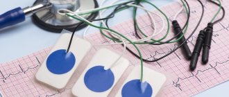
- electrocardiogram (ECG). With its help, a decrease in the ST segment and inversion of the T wave are recorded. The study allows us to identify the presence of arrhythmia, extrasystole and paroxysmal tachycardia;
- Magnetic resonance imaging (MRI) allows a reliable diagnosis to be made. Rarely used due to high cost;
- consultation with an endocrinologist and gynecologist.
As a result of a comprehensive study, it is possible not only to accurately establish the cause of myocardiostrophy, but also to choose the optimal tactics for its treatment.
Myocardial dystrophy ICD code 10. instructions for use
Meldonium is a structural analogue of gamma-butyrobetaine, a substance that is present in every cell of the human body. Meldonium suppresses gamma-butyrobetaine hydroxygenase, reduces the synthesis of carnitine and the transport of long-chain fatty acids through cell membranes, prevents the accumulation in cells of activated forms of unoxidized fatty acids - derivatives of acylcarnitine and acyl coenzyme A. Under conditions of ischemia, it restores the balance of the processes of oxygen delivery and its consumption in cells, prevents disturbances ATP transport. At the same time, it activates glycolysis, which occurs without additional oxygen consumption. As a result of a decrease in carnitine concentration, gamma-butyrobetaine, which has vasodilating properties, is intensively synthesized.
The mechanism of action determines the variety of its pharmacological effects: increased performance, reduced symptoms of mental and physical stress, activation of tissue and humoral immunity, cardioprotective effect. In case of acute ischemic damage to the myocardium, it slows down the formation of a necrosis zone and shortens the rehabilitation period. In heart failure, it improves myocardial contractility, increases exercise tolerance, and reduces the frequency of angina attacks. In acute and chronic ischemic disorders of cerebral circulation, it improves blood circulation in the area of ischemia. Effective in case of vascular pathology of the fundus. The drug eliminates functional disorders of the nervous system in patients with chronic alcoholism during the abstinence period.
complex therapy of coronary heart disease (angina pectoris, myocardial infarction); chronic heart failure, dyshormonal cardiomyopathy;
complex therapy of acute cerebrovascular accident (ischemic stroke, cerebrovascular insufficiency);
reduced performance; physical overexertion (including among athletes);
withdrawal syndrome in chronic alcoholism (in combination with specific therapy);
hemophthalmia and hemorrhages in the retina of various etiologies, thrombosis of the central retinal vein and its branches, retinopathy of various etiologies (diabetic, hypertensive).
age under 18 years (efficacy and safety have not been established).
The safety of the drug during pregnancy has not been proven. To avoid possible adverse effects on the fetus, the drug should not be prescribed during pregnancy. It is not known whether the drug is excreted in breast milk. If it is necessary to use the drug Idrinol® during lactation, breastfeeding should be discontinued.
Can be combined with antianginal drugs, anticoagulants, antiplatelet agents, antiarrhythmic drugs, diuretics, bronchodilators.
Enhances the effect of cardiac glycosides.
Due to the possible development of moderate tachycardia and arterial hypotension, caution should be exercised when combined with nitroglycerin, nifedipine, β-blockers, antihypertensive agents and peripheral vasodilators.
JSC "Pharm"
141345, Russia, Moscow region. Sergiev Posad municipal district, rural settlement Bereznyakovskoye, village. Belikovo 11.
Tel/fax
Owner of the registration certificate: CJSC Pharm.
Consumer complaints should be sent to the manufacturer's address.
On prescription.
Synonyms of nosological groups
Heading ICD-10 Synonyms of diseases according to ICD-10I25.9 Chronic ischemic heart disease, unspecified IHD Coronary atherosclerosis in patients with IHD Coronary circulatory insufficiency I21 Acute myocardial infarction Left ventricular infarction Myocardial infarction without Q wave Myocardial infarction in the acute period Non-transmural myocardial infarction (subendocardial) Acute myocardial infarction Myocardial infarction with and without pathological Q wave transmural myocardial infarction Myocardial infarction complicated by cardiogenic shock Non-transmural myocardial infarction Acute phase of myocardial infarction Acute myocardial infarction Subacute stage of myocardial infarction Subacute period of myocardial infarction Subendocardial myocardial infarction Coronary artery thrombosis (arthrombosis) terium) Threatening myocardial infarction I20 Angina pectoris [angina pectoris] Heberden's disease Angina pectoris Attack of angina Recurrent angina pectoris Spontaneous angina pectoris Stable angina pectoris Angina syndrome X Angina pectoris Angina pectoris (attack) Angina pectoris Angina pectoris Angina pectoris rest Angina progressive Angina mixed Angina spontaneous Angina stable Chronic stable angina I50.0 Congestive heart failure Anasarca cardiac Decompensated chronic heart failure Congestive circulatory failure Congestive heart failure with high afterload Congestive chronic heart failure Changes in liver function in heart failure Cardiomyopathy with severe chronic heart failure insufficiencyCompensated chronic heart failureSwelling due to circulatory failureSwelling of cardiac originSwelling of the heartEdematous syndrome in heart diseasesEdematous syndrome with congestive heart failure Edema syndrome with heart failure Edema syndrome with heart failure or cirrhosis of the liver Right ventricular failure Congestive heart failure Congestive heart failure Heart failure with low cardiac output Chronic heart failure Cardiac edema Chronic decompensated heart failure Chronic congestive heart failure Chronic heart failure I4 3.1 Cardiomyopathy in metabolic disorders Dishormonal cardiomyopathy Climacteric myocardial dystrophy I67.9 Cerebrovascular disease unspecifiedAngioneuropathyArterial angiopathyCerebral hypoxiaDyscirculatory encephalopathyCerebral disease of a vascular and age-related natureComa due to cerebrovascular accident Lacunar statusMetabolic and vascular disorders of the brainImpaired blood supply to the brainImpaired cerebral circulationImpaired functions of the brainImpaired functions of the cerebral cortexImpaired cerebral circulationInsufficiency of cerebral circulationAcute insufficiency cerebrovascular accident Acute cerebrovascular accident Damage to cerebral vessels Progression of destructive changes in the brain Cerebrovascular accident Syndrome cerebral insufficiency Vascular cerebrovascular insufficiency Vascular encephalopathy Vascular diseases of the brain Vascular disorders of the brain Vascular lesions of the brain Functional disorders of the brain Chronic cerebral ischemia Chronic circulatory insufficiency Chronic cerebrovascular insufficiency Chronic cerebrovascular insufficiency Chronic cerebral vascular insufficiency Cerebral insufficiency Cerebral organic failure Cerebral vascular insufficiency school pathologyCerebroasthenic syndromeCerebrovascular diseaseCerebrovascular diseaseCerebrovascular disorderCerebrovascular disorderDiscirculatory encephalopathyI63 Brain infarctionIschemic strokeIschemic brain diseaseIschemic brain lesionsIschemic strokeIschemic stroke and its consequences Ischemic cerebral stroke Ischemic cerebrovascular accident Ischemic brain damage Ischemic brain damage Ischemic condition Cerebral ischemia Acute cerebral hypoxia Acute cerebral ischemia Acute ischemic cerebrovascular accident Acute cerebral infarction Acute ischemic stroke Acute period of ischemic stroke Focal cerebral ischemia Post-existing ischemic stroke Repeated stroke Syndrome Morgagni-Adams-Stokes rumChronic cerebral ischemiaCerebrovascular strokeEmbolic strokeI67 Other cerebrovascular diseasesPain syndrome with vertebrogenic lesionsVertebro-basilar insufficiencyVertebrobasilar insufficiencyCerebral circulatory disordersAcute disturbance of blood supply to the brainCerebrovascular insufficiencyZ73.6 Limitations in activity caused by decreased ability to workGeneral weaknessMental fatigueSyndrome of decreased performance and physical overstrainWeaknessDecreased motor activityDecreased performanceDecreased physical performanceReduced performance Reduced performance of the body Reduced performance when working in extreme situations Z73.0 Overfatigue Restoration of physical performance of athletes Restoration of energy body statusGeneral weaknessMental fatigueE63 Physical and mental overloadRecovery period in athletesRestoration of physical performance of athletesHigh psycho-emotional stressHigh psycho-emotional stressHigh physical and mental stressHigh physical stressProlonged overload and stressProlonged physical and mental stressProlonged physical activityIntellectual stressIntense mental and physical stressIntense physical activityStrong mental activityImpaired mental work abilities Lack of nutrition due to physical and mental overload Nervous -emotional overload Nervous and physical overstrain Nervous overstrain Nervous overfatigue Increased mental and physical stress Increased mental stress Increased physical activity Increased physical and mental stress Increased physical and psycho-emotional stress Increased physical and emotional stress Increased physical stress Increased psycho-emotional stress Increased physical stress Decreased mental work abilities Decreased physical and mental performance Reduced performance Mental and physical fatigue Psychological and physical stress Psycho-emotional overload Psycho-emotional stress Psycho-emotional disorder Syndrome physical overstrain Decreased mental performance Reduced tolerance of psychophysical stress Reduced mental performance Fatigue state Mental and psycho-emotional stress Mental and physical stress Mental and physical overload Mental and physical fatigue Mental stress Mental and physical exhaustion Mental and physical stress Mental and physical stress Mental and physical fatigue Mental overload Increased physical and mental physical loadPhysical and mental stressPhysical and mental performancePhysical stressPhysical overloadPhysical and emotional overloadPhysical overloadPhysical stressPhysical and mental stressPhysical and mental stressPhysical overstrainPhysical stressChronic fatigueChronic physical overstrainExcessive physical and mental stressExcessive physical and mental stressExcessive physical stressExcessive mental and physical stressExtreme physical stressH44.8 Other diseases of the eyeballHemophthalmosEye adhesionsH35.6 Retinal hemorrhage bleeding Hemorrhagic retinopathy Retinal hemorrhage Retinal hemorrhage Hemorrhages in the eye Hemorrhages in retina at heights Repeated hemorrhage in the retina Mouth spots F10.3 Withdrawal condition Alcohol withdrawal syndrome Withdrawal syndrome Withdrawal syndrome in alcoholism Abstinence Alcohol withdrawal Alcohol withdrawal Alcohol withdrawal condition Alcohol withdrawal syndrome Post-withdrawal disorder Post-withdrawal condition Hangover syndrome Withdrawal syndrome tions Alcohol withdrawal syndrome Alcohol withdrawal syndrome Withdrawal condition H34 Retinal vascular occlusions Arterial thrombosis of the vessels of the eye Venous thrombosis of the vessels of the eye Retinal circulation disorders Intraocular circulatory disorders Insufficiency of blood supply to the retina and choroid eyes Occlusion of the central vessels of the retina Acute obstruction of the retinal arteries Subacute and chronic circulatory failure in the retina or in the choroid of the eye Vascular diseases of the retina Vascular disorders in the retina of the eye Thrombosis of the retinal vessels Thrombosis of the central retinal vein Thrombosis of the central retinal vein and its branches Thrombosis of the central retinal vein and its branches H35.0 Background retinopathy and retinal vascular changesAtherosclerotic retinopathyRetinopathyRetinopathyRetinopathy of newbornsH36.0 Diabetic retinopathy (E10-E14+ with a common fourth sign .3)Hemorrhagic diabetic retinopathyDiabetic retinopathyRetinal dystrophy in patients with diabetes mellitusH36.8 Other retinal disorders in diseases classified elsewhereAr theriosclerotic retinopathyAtherosclerotic retinopathyHypertensive retinopathy
Treatment and prognosis
Basically, the treatment of DCM comes down to symptomatic therapy, which involves taking:
- drugs that relieve cardialgia (Verapamil, Anaprilin);
- sedatives;
- vitamin complexes;
- immunostimulants;
- metabolic correctors (Actovegin, Mildronate).
If the above recommendations do not give a positive effect, hormonal treatment must be used. In this case, medications containing estrogens, gestagens or androgens are prescribed. The use of hormones is called “therapy of despair”, because even one dose of these substances can lead to disruption of the functioning of all endocrine glands of the body.
Therefore, when prescribing hormonal therapy for DCM, a number of factors must be taken into account:
- therapy should be carried out in long-term cycles and only under the supervision of a physician;
- dose selection is individual in each specific case;
- the effectiveness of treatment is judged by the patient’s condition, and not based on the results of examinations, which are always somewhat delayed.
Non-drug therapy includes:
- complete elimination of bad habits;
- playing sports;
- dietary nutrition;
- balanced daily routine;
- alternation of work and rest.
Psychological support is another integral component of DCM treatment. The patient should be informed that his condition is not critical and not life-threatening.
Forecast
In most cases, the prognosis for DCM is favorable. The patient must be explained that the pain syndrome does not threaten his life and is not associated with cardiac activity. This disease does not require bed rest; on the contrary, you need to lead a normal active lifestyle for a speedy recovery. In most cases, after endocrine changes are completed, all negative manifestations are eliminated on their own. If this does not happen, then correctly selected treatment prescribed by a competent specialist will help get rid of annoying symptoms and avoid unpleasant consequences.
conclusions
DCM is a cardiac disorder that occurs due to dysfunction of the endocrine system. The disease can occur during menopause and premenstrual periods, due to thyrotoxicosis, as well as other conditions with hormonal imbalance in the human body. All this leads to the appearance of structural and functional changes in the heart, which will be manifested by corresponding symptoms. Treatment of this disease is conservative and is aimed at eliminating the factor leading to dystrophic disorders. To avoid negative consequences, it is necessary to avoid self-medication and consult a doctor at the first symptoms.
Methods for diagnosing functional cardiopathy
Clinical and instrumental methods are used for diagnosis
ECG: signs of myocardial hypertrophy, rhythm and conduction disturbances, ST changes.
X-ray of the lungs: you can see hypertrophy, dilatation of the myocardium, congestion in the lungs.
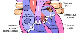
EchoGK: allows you to evaluate the size of the heart cavities, the condition of the valves, the thickness of the walls and interventricular septum, and evaluate systolic and diastolic functions.
Sometimes they use: MRI, radioisotope ventriculography, angiocardiography, cardiac catheterization, endomyocardial biopsy is taken.