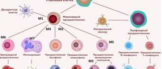
How to decipher the leukocyte formula?
Leukocyte formula, microscopy of leukocytes, five fractions of leukocytes, differentiation of leukocytes - in doctor’s prescriptions you can find many names for the same thing. Where can I find it and how to decrypt it?
What are leukocytes?
Leukocytes (white blood cells) are a large group of blood cells. Their main purpose is to protect the body from infections. All leukocytes are part of the immune system; they are involved in allergic, autoimmune, and tumor processes. Each type of white blood cell has its own role and is important for the body.
A general blood test without a leukocyte formula speaks only about the total content of leukocytes and does not reveal what type of leukocytes is responsible for the increase (leukocytosis) or decrease (leukopenia) of white blood cells. The leukocyte formula determines five types of leukocytes and is assessed in a comprehensive general blood test. To decipher the leukocyte formula, you need to evaluate the content of each type of leukocyte and their ratio to each other.
The leukocyte formula is calculated by an automatic blood analyzer. Taking the content of all white blood cells as 100%, it gives the percentage (%) of each type of white blood cell. It also automatically measures their content in blood volume (per liter). Sometimes a “hand count” and visual assessment of the blood smear under a microscope is required. For example, when the white blood cell count is altered, there are strange or immature cells, there are signs of anemia or a decrease in platelets in the general blood test. In this case, you can only see the percentage of the leukocyte formula.
Granulocytes - shock forces
The largest part of leukocytes is represented by granulocyte cells. They got their name due to the presence of inclusions (granules). The granules contain immune chemicals. In the leukocyte formula you can see three types of granulocytes: neutrophils, eosinophils, basophils. They differ in the structural features of the core and the coloring of the granules with different dyes. Granulocytes are important in the development of inflammation and immune defense of the body. They are capable of absorbing and digesting proteins and chemicals. All granulocytes mature in the bone marrow, maintaining a supply of mature cells there for 3-4 days. Granulocytes circulate in the blood for no more than 6 hours, moving into the tissues where they perform their function.
Neutrophils make up the largest number of leukocytes circulating in the blood. Every day, 1010 neutrophils enter the bloodstream. Neutrophilia (an increase in the number of neutrophils in the blood) is an indicator of a bacterial infection. The more severe the infection, the more neutrophils come out to fight. Due to their low life expectancy (about 4 days), younger, immature forms of cells (band cells, metamyelocytes, and others) begin to enter the blood. Doctors call this a “leukocyte shift to the left.” When there are very few neutrophils in the blood (neutropenia), the body is not protected from infections.
Eosinophils in the blood make up no more than 5% of the total number of leukocytes. Their concentration fluctuates throughout the day due to the influence of adrenal hormones. In the morning it is maximum. They accumulate in the submucosal layer of the gastrointestinal tract. Eosinophilia (increased eosinophilia in the blood) occurs with parasitic infections, allergic and autoimmune processes.
Basophils make up the smallest number of leukocytes in the blood (less than 1%) and are involved in allergic reactions by releasing histamine. This substance is responsible for bronchospasm, itching, swelling, and redness. Depending on where the basophils get to, there will be manifestations of allergic reactions: an attack of bronchial asthma, skin rash, urticaria, Quincke's edema (swelling of the larynx).
Monocytes are tissue hunters
The second link of leukocytes is monocytes. In the bone marrow, having formed in 5 days, they do not form a reserve. In the blood, monocytes make up about 10% of the mass of leukocytes, quickly disappearing into the tissues. Tissue macrophages, and this is what monocytes will be called, are mainly found in the liver, spleen, and lungs. Their life expectancy is very long (60 days). They are the main hunters of the immune system, because... absorb and process thousands of foreign proteins, turning them into antigens accessible to immune cells.
Monocytosis (an increase in monocytes in the blood) is associated with chronic infections, as well as with infections whose pathogens are hidden in the body’s cells (viruses, chlamydia, mycoplasma).
Lymphocytes are reliable defenders
Antigens processed by monocytes-macrophages and other immune cells attract lymphocytes. Lymphocytes provide acquired immunity by producing antibodies and memory cells to protect against re-infection.
Lymphocytes are formed in the bone marrow and circulate both in the blood and in the lymphatic system. Important organs of cell maturation are the thymus (thymus gland) and lymph nodes. Lymphocytes perform various immune functions, representing the second largest group of leukocytes. There is a special blood test (immunophenotyping) that allows you to identify different types of lymphocytes. This can be important for diseases of the immune system, HIV infection, etc.
In the leukocyte formula, both an increase in lymphocytes (lymphocytosis) - more typical for viral infections - and lymphopenia (a decrease in their number) are important. A lack of lymphocytes reduces the body's defenses and is observed in immunodeficiencies (including HIV infection).
In the Lab4U laboratory you can take the following tests with a 50% discount:
- General blood test with leukocyte formula (automatic counting)
- General blood test with microscopy (manual leukocyte count)
Norms of myelocytes in the bone marrow
Blood cell maturation
Since immature forms of leukocytes must undergo all stages of maturation in the bone marrow, they should normally be present only in the bone marrow puncture.
If a person is healthy, then there is no reason for immature forms of blood cells to enter the systemic circulation.
To assess the consistency of granulocytopoiesis and all hematopoiesis in general, a research method such as bone marrow puncture (sternal puncture, trephine biopsy) is used.
Normally, the granulocytic (myelocytic) lineage of hematopoiesis will give the following indicators when assessing the myelogram:
| Cellular composition of the bone marrow (granulocytopoiesis) | Quantity, % |
| Undifferentiated blast cells | 0,1-1,1 |
| Myeloblasts | 0,2-1,7 |
| Promyelocytes | 1,0-4,1 |
| Myelocytes | 6,9-12,2 |
| Metamyelocytes | 8,0-14,9 |
| Rod | 12,8-23,7 |
| Segmented | 13,1-24,1 |
| Neutrophil Maturation Index | 0,5-0,9 |
| All eosinophils | 0,5-5,8 |
| Basophils | 0-0,5 |
Myelocytes in children and pregnant women
Myelocytes can appear when the immune defense in children is reduced
The detection in the blood of any cell that has not completely completed its differentiation during granulocytopoiesis indicates that the bone marrow has become activated in response to some pathological transformation.
In a healthy child, myelocytes, like other young forms, should not be detected in the blood. The release of myelocytes into the blood, as well as other maturing granulocytes, is determined by the same factors as in adults. Also, the detection of immature cells in babies is often diagnosed with congenital heart defects, uncontrollable vomiting and dehydration. Very often, immature forms of granulocytes are found in children with weak immunity.
Severe physical stress will also lead to the appearance of a small number of myelocytes in the blood of healthy children.
As for pregnant women, fluctuations in the blood picture are allowed. The process of hematopoiesis during pregnancy is enhanced to maintain the vital functions of the entire body of the mother and baby. Additionally, the appearance of myelocytes and other young forms in the blood may be the result of an exacerbation of chronic diseases, for example, sinusitis, pyelonephritis, etc.
In expectant mothers, the concentration of myelocytes in the blood is allowed to be no more than 2-3%. But, in any case, this phenomenon requires further diagnosis in order not to miss the development of malignant pathology.
“Illegal” penetration into peripheral blood
However, there are situations when cells that still need to “grow and develop” prematurely leave their “native homes”. And if normally the appearance of blast cells in the peripheral blood is out of the question - they are rare “guests” in the bloodstream, then under certain pathological conditions, contrary to natural prohibition, both of them still enter the bloodstream.
Blasts and myeloblasts are slightly increased (up to 2% of the total leukocyte population) in chronic forms of leukemia . And a huge number of blasts (blastemia) generally indicates serious changes in the hematopoietic organs and is one of the significant signs of acute leukemia, the form of which will subsequently be clarified by other methods.
Of particular concern is the number of blasts crossing the 5% limit in the blood of a patient suffering from chronic myeloid leukemia - this may indicate the onset of a blast crisis and the final stage of the tumor process.
myeloblasts in the blood
The presence of propromyelocytes, myelocytes and the closest to mature forms - metamyelocytes, although not such a terrible indicator of white blood, still indicate a serious pathology. An increase in the number of these cells to 5% is more often caused by non-hematological pathology:
- A severe infectious disease of any origin: both bacterial (mainly) and viral;
- Development of a septic condition;
- Various types of intoxication (bacterial, alcoholic, salts of heavy metals);
- Tumor (malignant) process;
- Chemotherapy and radiation therapy;
- Taking certain medications (analgesics, immunomodulators);
- Acute blood loss;
- Coma, shock;
- Violation of acid-base balance;
- Excessive physical activity.
presence of myelocytes and metamyelocytes in the blood
Meanwhile, a significant jump in myelocytes, pro- and meta- (up to 10–25%), is usually observed in the case of the formation of myeloproliferative diseases, which are the most basic reasons for the exit of maturing forms from the bone marrow and their free movement through the blood vessels.
“Young and early”...
The collective name “myeloproliferative tumors” refers to chronic leukemias that are formed at the level of the youngest precursors of myelopoiesis, all of whose offspring - granulocytes, monocytes, erythrokaryocytes, megakaryocytes (except lymphocytes) belong to the tumor clone.
Chronic myeloid leukemia, opening the list of myeloproliferative processes, acts as a typical representative of tumors that arise from early (very young) precursors that differentiate through myelopoiesis to a mature state.
The cellular substrate of myeloid leukemia originates from the white germ of hematopoiesis and is represented by transitional (maturing) forms of granulocytes, mainly neutrophils. This suggests that important cells such as neutrophils, which play such an important role in protecting the body, suffer the most, which makes it clear why this disease is so difficult to treat and ultimately fatal.
At the beginning of the disease, a shift to myelocytes and promyelocytes is noted in the blood, although their number at first is still insignificant. In addition to single promyelocytes and a slightly larger number of myelocytes, representatives of other cell populations can be found in the blood (erythrokaryocytes, counted in units, and high thrombocytosis).
The advanced stage of the disease gives a significant rejuvenation of the leukocyte formula and, at the same time, in addition to myelocytes, the absolute values and percentage of already mature forms of the granulocyte series in the blood are often increased: eosinophils or basophils (less often, both of them - “basophilic-eosinophilic association”). It should be noted that a sharp increase in the number of immature neutrophils is a very, very unfavorable sign that complicates the course of the disease and prognosis.
Treatment
Elimination of the cause leads to normalization of the blood picture
Since myelocythemia is caused by the development of a disease, it is the root cause that requires treatment.
Only after the cause of the pathological change in blood composition has been clarified, treatment is prescribed.
There are many reasons for the penetration of immature blood cells, in particular myelocytes. The vast majority of such triggers, unfortunately, are dangerous diseases that can quickly claim a human life. At the slightest transformation in the composition of the blood, it is necessary to urgently consult a doctor, diagnose and prescribe the necessary treatment methods.
How to determine myelocyte level
A myelogram will show the exact level of myelocytes
Determination of the level of myelocytes, as well as other components of the bone marrow, is carried out by taking a bone marrow puncture. The resulting myelogram will show the exact cellular composition of the bone marrow.
Bone marrow samples are taken from:
- sternum,
- iliac crest,
- from the calcaneus (this is how puncture is performed in small children).
After receiving the punctate, the myelogram data must be compared with the data of the general clinical blood test.


