When the heart rate increases, a diagnosis of sinus tachycardia is often made. What can it mean, especially if the disorder is detected in children of different ages? Most often, a change in rhythm does not pose any particular danger, but it is additionally necessary to know when medical attention may be required.
Sinus tachycardia (ST) is a change in heart rhythm towards its increase, in which the sinus node remains the main pacemaker. In children, this condition is very common, which is often associated with both their increased activity and fairly rapid growth.
In pathological cases, tachycardia is observed in a calm state, and in addition to rapid heartbeat, other pathological signs are determined.
Tachycardia is diagnosed simply, for which it is enough to correctly count the heart rate in the radial or carotid artery. To determine the type of tachycardia, electrocardiography will be required. If the rhythm disturbance does not affect the child’s well-being, then treatment is not carried out. Otherwise, additional diagnostics are performed and then appropriate therapy is prescribed.
Video: Sinus tachycardia in a child
Causes of tachycardia in children
Do not forget that even normally, the heart rate (HR) of a child is significantly higher than that of an adult. This is due to a more active metabolism, leading to increased oxygen consumption by the cells of a growing organism. Heart rate norms for children of different ages are indicated in the table below:
| Child's age | Average heart rate (beats/minute) |
| Newborn up to 2 days | 123 |
| 2 – 6 days | 129 |
| 7 – 30 days | 148 |
| 30 – 60 days | 149 |
| 3 – 5 months | 141 |
| From six months to 11 months | 134 |
| 12 – 24 months | 119 |
| 34 years | 108 |
| 5 – 7 years | 100 |
| 8 – 11 years | 91 |
| 12 – 15 years | 85 |
| Over 16 years old | 80 |
The causes of tachycardia in children can be both physiological and pathological; associated with heart disease or resulting from other ailments.
With a healthy cardiovascular system, an increase in heart rate can be triggered by the following factors:
- hyperthermia due to acute respiratory viral infections or other infectious diseases;
- high ambient temperature;
- increased physical activity;
- emotional overexcitation;
- thyroid diseases;
- vegetative-vascular dystonia;
- overweight;
- pheochromocytoma;
- dehydration;
- anemia.
As for heart diseases, tachycardia in children is most often observed with congenital heart defects, myocarditis, and certain types of conduction disorders. Depending on the age of the child, one or another reason comes first.
In infants
Babies in the first months of life are most often exposed to attacks of tachycardia under the influence of the following factors:
- external mechanical influences, such as inspections or changing;
- cardiovascular failure;
- congenital developmental anomalies;
- perinatal encephalopathy;
- respiratory failure;
- acute asphyxia;
- hypoglycemia;
- anemia.
Naturally, a baby is not immune from the development of infectious diseases and, first of all, colds.
For schoolchildren
The reasons why tachycardia may develop in a child who has begun to attend school differ from those in an infant. By this time, congenital developmental anomalies partially lose their positions, since they have been cured, stabilized, or have already led to a sadder result, but continue to remain in first place. The frequency of acute asphyxia is also noticeably reduced. The main reasons for high heart rate in schoolchildren are:
- autonomic disorders due to increased stress, both psychological and physical;
- thyroid dysfunction;
- hyperthermia of various origins;
- organic heart pathologies;
- excessive physical activity;
- electrolyte dysfunctions;
- neoplasms.
The task of parents of schoolchildren who want to reduce the likelihood of tachycardia in their children is to normalize the regime, minimize physical and psychological stress on the child, undergo regular medical examinations and fight against infectious pathologies that can cause inflammation of the membranes of the heart.
All carious teeth need to be cured, and if a child has a sore throat or already feels discomfort in the heart due to an elevated temperature, then no exam or new topic should be a reason to postpone a visit to the doctor.
In teenagers
Adolescence is characterized by a significant increase in the development of risks for the cardiovascular system. This time marks the beginning of increased body growth, puberty with a characteristic surge of emotions, and the first experiments with the use of psychoactive substances.
Let's figure out what reasons cause rapid heartbeat in a teenager.
Their list is as follows:
- chronic infections (caries, frequent sore throats) that provoke inflammation of heart tissue;
- imbalance in growth rate between the heart (lags behind) and the rest of the body (leads);
- general passion for tonic low-alcohol drinks;
- “small heart” against the background of an underdeveloped musculoskeletal system;
- vegetative-vascular manifestations;
- electrolyte imbalance;
- tumors.
From this list, parents, even without contacting doctors, can cope with a good third of the reasons.
Boys and girls should be taught the negative effects of drinks containing caffeine and other stimulants. If he or she really wants alcohol before a disco, let him or her drink wine or vodka rather than Red Bull or Jaguar.
Diagnostics
During the examination of the child, the doctor may ask the following questions:
- Does the child have a medical history, particularly sinus tachycardia, or any other heart condition?
- What was the onset and duration of the child’s current illness?
- Does the child have other symptoms, such as:
- chest pain;
- shortness of breath;
- change in skin color;
- loss of consciousness;
- neurological symptoms (eg, changes in level of consciousness);
- Is the child taking any medications?
- Does the child have an allergy to anything?
- Is there a family history of heart disease?
- Does tachycardia occur at a specific time or under certain circumstances (eg, during activity, after eating, under stress)?
- Has the child complained of feeling hot/fever?
- Has the child had vomiting or diarrhea?
- How often does your child drink and urinate?
In addition to a complete medical history and physical examination of the child, there are various types of procedures that may be used to diagnose TS. In particular, the following methods are used:
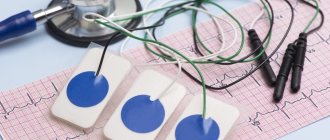
- Electrocardiography (ECG) is a measurement of the electrical activity of the heart. When electrodes are placed in certain places on the body (chest, arms and legs), a graphic image is created in the form of teeth of various heights and shapes. An ECG may indicate the presence of arrhythmia or other types of heart disease.
There are various options for an ECG test, including:
- Resting ECG (resting ECG) - to conduct this study, the child undresses to the waist, and then the doctor attaches electrodes in the form of small pads to the chest, shoulders and legs. On the other side, the electrodes are connected to an ECG machine. The device starts up and records the electrical activity of the heart for a minute or so. During this, the child lies quietly.
- Stress test . The child is connected to the measuring equipment as described above. However, instead of lying down, the child does the exercise by moving on a treadmill or “riding” a stationary bicycle. During the movement, an ECG is recorded. This test is carried out to assess the functioning of the heart during physical activity, which partly corresponds to the level of emotional stress.
- High resolution ECG . This procedure is performed in the same way as a resting ECG, except that the electrical activity of the heart is recorded over a longer period of time, usually 15 to 20 minutes. The method is used when arrhythmia is suspected, which is not recorded by a standard ECG, since rhythm disturbances can be short-term in nature and do not occur during recording in the usual way.
- Holter monitoring is an ECG recording that is performed over a period of 24 hours or more. To conduct the test, three electrodes are attached to the child's chest and connected to a small, portable ECG recorder using wires. The child goes about his or her normal daily activities (except for activities such as showering, swimming, or any activity that causes excessive sweating, which could cause the electrodes to fall off). There are two types of Holter monitoring: Continuous recording - the ECG is recorded continuously throughout the testing period.
- Event Monitor - An ECG is recorded only when the patient experiences a palpitations.
Differential diagnosis of sinus tachycardia with other types of arrhythmia
| Sinus tachycardia | Supraventricular tachycardia (re-entry) | Atrial fibrillation | |
| Clinic | May be absent or fever/sepsis/shock/anemia | 50% - Wolff-Parkinson-White syndrome 25% - atrioventricular, nodal, reentral tachycardia, Ebstein's anomaly | 90% have atrial dilatation/myocarditis/digoxin toxicity |
| Heart rate | Typically <200/min | Infants: <300/min, older children: <240/min. | Atrial rate 250-400/min 2:1, 3:1 or 4:1 with AV block |
| P wave | Normal | May be hidden in the QRS complex, 60% have retrograde P-wave | Regular flutter waves (F-waves) |
| QRS | Normal | Normal or aberrant | Normal |
Thus, sinus tachycardia has the most clinically favorable course, since other forms of arrhythmia are often accompanied by more pronounced and severe clinical symptoms.
What tachycardias are most common in children?
In a child, palpitations are divided into two main types:
- sinus;
- paroxysmal.
The first type occurs most often and, as a rule, against the background of a healthy heart.
Paroxysmal tachycardia is a whole group of diseases characterized by:
- sudden onset;
- high heart rate;
- spontaneous restoration of normal heart rhythm;
- preservation of the normal sequence of cardiac complexes on the ECG;
- short duration of the attack, from a few seconds to several days.
Frequency of occurrence in the pediatric population: 1 in 25,000 people, which is an average of 15% of all arrhythmias. Paroxysmal tachycardia is divided into the following forms:
- atrial;
- ventricular;
- atrioventricular.
Pathology develops as a consequence of such factors:
- congenital or acquired during childbirth pathology of the central nervous system;
- unfavorable family and social situation;
- congenital malformations of the child’s heart;
- cardiac surgery;
- some infectious pathologies;
- installation of a catheter in the heart cavity;
- heart trauma (closed);
- angiocardiography.
Another attack can be triggered by:
- mental stress;
- physical overload;
- hyperthermia;
- stress.
A child with paroxysmal tachycardia presents the following complaints:
- palpitations starting with a “push” behind the sternum;
- pain in the area of the heart and in the pit of the stomach;
- feeling of lack of air;
- dizziness;
- headaches;
- insomnia;
- weakness;
- nausea;
- fear.
As for changes on the ECG, they differ depending on the form of the disease and are indicated in the table below:
| Form of paroxysmal tachycardia | Changes on the cardiogram |
| Supraventricular | The P wave is associated with an unchanged QRS complex or is not detected, and can also have a wide variety of shapes. A sequence of extrasystoles of atrial origin is observed. Heart rate is from 160 beats/minute. |
| Ventricular | Short (five or more) sequences of extrasystoles of ventricular origin, alternating with short-term sinus intervals. The QRS complex is deformed, widened to 0.1 seconds or more. The P wave most often overlaps with other elements and is therefore almost never detected. |
This disease can be life-threatening for the child and requires emergency treatment.
Sinus tachyarrhythmia in a child
This type occurs due to increased functioning of the sinus node. This condition can be provoked by a number of irritants:
- stress;
- dehydration;
- shock states;
- physical activity;
- an increase in the concentration of calcium ions in the blood;
- consumption of large doses of stimulants (tea, coffee);
- taking medications (caffeine, antidepressants, antiallergic drugs, theophylline and some others).
The main external distinctive signs of sinus tachyarrhythmia are:
- short duration;
- no significant discomfort;
- normalization of heart rate after removal of the influence of the irritating factor.
Tachycardia, which persists for a long time, can develop under the influence of various pathological conditions associated with both heart problems and diseases of other organs - anemia, respiratory failure, etc. In this case, an increase in heart rate is accompanied by certain complaints of moderate intensity: palpitations, sensation lack of air.
Sinus tachyarrhythmia is a condition characterized by a heart rate that exceeds the child’s age norm. It is based on the acceleration of the production of electrical impulses by the first-order pacemaker, the sinus node. In the absence of other symptoms, in addition to an increase in heart rate, sinus tachycardia is considered a normal variant.
With a significant acceleration of the heart, the child experiences the following symptoms:
- fatigue and weakness inappropriate for physical activity;
- emotional arousal;
- change in skin color;
- dizziness;
- heartbeat;
- moodiness;
- fussiness and so on.
Sinus tachycardia in most cases resolves spontaneously immediately after the influence of the provoking factor ceases.
To make a diagnosis of sinus tachycardia, the following methods are used:
- taking anamnesis;
- physical examination;
- types of ECG (regular, stress test, high resolution, Holter monitoring);
- electrophysiological study.
Differential diagnosis with other rhythm disturbances is of great importance. Sinus tachycardia of all arrhythmias has the most favorable prognosis.
Description of sinus tachycardia
Sinus tachycardia is one of the types of arrhythmias in which the heart rate is higher than typical for the child’s particular age. This is due to changes in the sinus node, which sends electrical impulses at a faster rate than normal.
In a normal state, the heart rhythm is set by the first-order driver - the sinus node, therefore, if only one indicator (heart rate) changes and there are no clinical manifestations, sinus tachycardia is defined as a variant of the norm.
If the heart rate becomes too fast, the body's circulatory process is disrupted. In such cases, TS may cause the following symptoms:
- weakness;
- fatigue;
- dizziness;
- feeling of heartbeat.
In young children, TS can manifest as fussiness, difficulty feeding, moodiness, excessive agitation, changes in skin color, etc.
Sinus tachycardia is often temporary when the body is under stress from exercise, strong emotions, fever, or dehydration. Sometimes a child is affected by several reasons at once. Once the external factors are eliminated, the heart rate usually returns to normal.
How to act
When any type of arrhythmia appears in a child, parents are required to first call an ambulance. And only after the call or in parallel with it, begin to provide first aid:
- unfasten tight clothing on the child’s chest and neck;
- provide fresh air access to the room;
- Place a damp cloth on the patient's forehead.
It would be nice to try the so-called vagal tests:
- turn the baby upside down for half a minute; an older child can be helped to stand in his arms for the same time;
- ask him to tense his abdominals, strain, while holding his breath, press the child on the epigastrium (these actions are also performed for 30 - 40 seconds);
- press the root of the tongue and induce vomiting;
- Immerse the child’s face in a bath of cold water (procedure duration is from 10 to 30 seconds).
It is clear that these actions can only be performed with a child over 7-10 years old, to whom the meaning of the manipulations can be explained.
I would not recommend another test, which requires massaging the carotid sinus, without special preparation, since it is necessary to press the carotid artery.
These tests can bring a positive effect within half an hour from the onset of an attack of tachycardia.
When to see a doctor and how often to get checked
You should consult a doctor immediately after parents notice any of the above symptoms of any tachyarrhythmia. And it would be better to play it safe and bother the pediatrician in case of physiological tachycardia, which developed in response to stress or physical overload, than to miss the first “bells” indicating the onset of the development of a serious disease.
All children are covered by regular medical examinations from the moment of birth, so identifying arrhythmias should not be difficult. However, unfortunately, it is not always possible to detect symptoms and prescribe treatment for tachycardia in children in a timely manner.
There are several reasons for this:
- the formal attitude of pediatricians towards mass examinations of children;
- parents' inattention to children's complaints;
- Children's fear of doctors, which is why they do not tell their parents and doctors about their problems.
The solution is simple: attention to your own child and regular ECG diagnostics, especially during attacks.
A lot depends on parents regarding early diagnosis. After all, doctors, unfortunately, are not psychics and do not sense from a distance when the baby will develop the first attack of tachyarrhythmia in his life, but dad and mom are quite capable of noticing this and contacting a doctor in time.
An electrocardiogram is the most revealing method for detecting arrhythmias. The difference between sinus and paroxysmal tachyarrhythmias is indicated in the pictures below:
In what cases is treatment required?
Only a doctor should decide whether treatment is necessary in each specific case and what it will consist of! Self-medication for tachyarrhythmias can end very sadly. Prescriptions are made by a pediatric cardiologist or, in non-critical cases, by a pediatrician after consultation with a cardiologist. Treatment is carried out in accordance with approved protocols and can be either therapeutic or surgical.
Functional rhythm disturbances do not require treatment; it is enough to organize the child’s correct schedule of work, study, and rest.
A comprehensive approach should be taken to clinically significant arrhythmias. Therapy should begin with the removal of all chronic infectious foci and treatment of diagnosed rheumatism.
In the conservative treatment of childhood tachyarrhythmias, there are three main directions:
- bringing the electrolyte balance in the heart muscle to normal levels (preparations of magnesium and potassium ions);
- taking antiarrhythmic drugs (Verapamil, Propranolol, Amiodarone, etc.);
- improvement of metabolism in the myocardium (Riboxin, Cocarboxylase).
If rhythm disturbances are resistant to the action of drugs, then the turn comes to minimally invasive surgical interventions:
- radiofrequency or cryoablation of arrhythmogenic foci;
- implantation of a cardioverter-defibrillator or pacemaker.
In the vast majority of cases, arrhythmias that developed in childhood, subject to timely consultation with a doctor, are completely cured or stabilized.
Treatment
Specific treatment for sinus tachycardia will be determined by the child's physician based on:
- Age, general health and medical history
- Degrees of clinical manifestations
- Tolerance of the child's body to specific medications, procedures or therapies
Sinus tachycardia may be present but not noticeable or cause any health problems. In this case, the doctor can only recommend adjusting your lifestyle, if necessary. However, when a rhythm disorder causes symptoms, there are several different treatment options.
As a rule, the doctor chooses treatment tactics according to the type of arrhythmia, the severity of symptoms, and the presence of other pathological conditions (for example, diabetes, kidney failure, heart failure, thyroid dysfunction) that may affect the course of treatment.
Most often, sinus tachycardia can be treated with lifestyle changes and medications. In other cases, the following therapy methods are used:
- Cardioversion . This procedure uses a low electrical voltage delivered to the heart through the chest to stop certain very rapid arrhythmias, such as atrial fibrillation, supraventricular tachycardia, or sinus tachycardia. The child is first given medication to help him relax and is then connected to an ECG monitor that works in parallel with a cardioversion device. A weak electrical current is applied to a specific point in the heart during an ECG cycle.
- Ablation is an invasive procedure performed in the electrophysical therapy department. It is carried out through a small thin tube (catheter) that is inserted into the heart through a vessel in the groin or arm. The procedure is carried out similarly to the electrophysiological study described above. Once the causative site of the arrhythmia is determined using EPI, the catheter is moved to it. The cause of the rhythm disorder can be destroyed using the appropriate technique: Radiofrequency ablation (the affected area is exposed to very high-frequency radio waves, which heat the tissue until it is destroyed)
- Cryoablation (part of the myocardium is exposed to an ultra-cold substance that freezes the tissue and then destroys it).
In infants and young children, pacemakers are usually placed in the abdomen. The wires that connect the pacemaker to the heart are placed on the outer surface of the heart. This precaution is necessary because fat in the abdomen protects the pacemaker wires and the device from shocks that may occur during daily childhood activities, such as standing up or falling.
For school-age children and teenagers, a pacemaker may be placed in the shoulder area under the collarbone. Pacemaker wires are often placed inside the superior vena cava, a large vein that connects to the right atrium, and then guided into the heart.
Surgery is usually performed only when all other modern therapies have failed to produce the expected results. Surgical ablation is a major surgical procedure requiring general anesthesia. The chest is cut open and open heart surgery is performed. The location of the arrhythmic focus is determined, then it is destroyed or removed, which eliminates the arrhythmia.
Video: What is cardiac tachycardia and how to treat it?
Should parents worry?
Regardless of whether the child has complaints, parents should be attentive to the state of his health. Indeed, in the life of a growing organism there are 4 periods of risk for the appearance of arrhythmias, through which everyone goes through:
- newborn;
- from four to five years;
- from seven years to eight;
- from twelve to thirteen years old.
Children of these age groups must undergo mandatory electrocardiographic examination. If a child presents even one cardiogenic complaint, then doctors should prescribe additional types of ECG, various tests and examinations.
If problems are identified, then you need to start treating the child without delay. Most arrhythmias have a favorable prognosis. Clear recommendations on treatment tactics have been developed; in severe cases, surgical interventions are performed. Arrhythmia is not a death sentence; it must be fought and can be defeated, returning the child to a full life.
Case from practice
I bring to your attention a very illustrative case where a combination of a number of unpleasant circumstances and mistakes led to serious health problems for a young girl.
Thirteen-year-old K. was sent to a specialized cardiac surgery hospital with complaints of pulling and stabbing pain behind the sternum on the left, not periodic, not associated with the emotional state, physical activity and changes in body position. Painful attacks passed after taking sedatives or on their own. She had been noticing the sensations for a couple of years and came in due to the worsening of her condition.
Anamnesis of life
She was born full-term from the second pregnancy. There are no hereditary complications, bad habits, or professional hazards on the part of the parents. In the first half of pregnancy, the mother suffered from severe toxicosis. No examination for intrauterine infections was carried out.
As she grew older, she suffered from the following diseases:
- 1 year - no pathologies;
- 4 years - scarlet fever;
- 5 years - HES;
- 6 years old - lacunar tonsillitis.
At an older age, I occasionally suffered from ARVI.
Electrocardiography was not performed until the age of ten!
An ENT doctor diagnosed and observed chronic tonsillitis in the compensation stage.
Physically developed in accordance with age standards, harmoniously.
History of the disease
For the first time I felt pain in my heart when I reached the age of ten and addressed these complaints to a cardiorheumatologist.
A month later, the pain returned and began to appear more often. Extrasystole and atrial flutter appeared. This time K. was hospitalized and she was prescribed antiarrhythmic drugs, which, however, did not bring the expected effect. The girl was sent to a cardiac surgery hospital.
The ECG shows sinus arrhythmia, and the EchoCG shows enlargement of the left ventricle. Mitral valve prolapse was also detected, which at that time had not yet led to hemodynamic disturbances. The attending physician prescribed daily ECG monitoring, with the help of which attacks of atrial arrhythmia were detected.
The prescribed drug antiarrhythmic therapy was effective and led to a decrease in heart rate during attacks of atrial arrhythmia and the cessation of relapses of atrial fibrillation.
The child was observed in the clinic for six months. Despite supportive treatment, atrial tachyarrhythmia persisted. Echocardiography revealed enlargement of the right chambers of the heart. Considering that conservative treatment methods failed to achieve either a complete cure or even reliable stabilization of the condition, and the conduction system of the heart was under constant threat of inflammatory changes, it was decided to perform a biopsy of endomyocardial tissue using an intracardiac catheter.
During the examination of the biopsy specimen, tissue degeneration and the presence of a large number of leukocytes were revealed.
Subsequently, the girl was treated with cardioprotectors, antiarrhythmics, and anticoagulants.
Sluggish chronic tonsillitis was treated in order to eliminate the source of pathogenic microflora in the body.
After the entire complex of measures taken, K.’s well-being improved, relapses of arrhythmias were not observed, and the size of the heart returned to normal. However, there is dysfunction of the sinus node, and the patient is being monitored.
Conclusion
The patient came to this difficult situation for several reasons:
- lack of ECG control during periods of life that threaten the development of arrhythmias;
- the presence of an unsanitized chronic source of infection in the body;
- latent, sluggish, difficult to diagnose course of endomyocarditis.
To prevent such cases in other children, you should be more attentive to preventive examinations, observing the principle of overdiagnosis. In doubtful cases, it is better to refer the child for additional examination, which will show the norm, than to miss the pathology!
Doctor's advice
In conclusion, I want to give some simple advice to those parents who want to minimize the risk of developing arrhythmias in their child:
- do not hesitate to ask the doctor for a referral for an ECG during the risky periods of the child’s life listed above;
- interpretation of children's ECGs from periodic examinations should be carried out in the department of functional diagnostics;
- sanitize all foci of chronic infection - carious teeth, diseases of the ENT organs, respiratory tract, skin;
- the child should sleep enough, eat well, drink plenty of fluids, and avoid eating synthetic and GMO foods;
- he should, if possible, be protected from unnecessary stress and heavy physical activity;
- At the slightest complaint, the child should be shown to specialists.
And remember that any disease, especially arrhythmia, is much easier to prevent than to treat.
