© Author: A. Olesya Valerievna, candidate of medical sciences, practicing physician, teacher at a medical university, especially for SosudInfo.ru (about the authors)
During operation, the heart produces many sound effects that characterize the process of myocardial contraction, valve movement, and the impact of moving blood on the walls of blood vessels. To register and evaluate these sounds, FCG - phonocardiography is used.
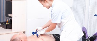
The most accessible way to determine sounds is auscultation, when the doctor listens to the heart using a phonendoscope. However, not all sound waves can be caught by the ear, even “armed” with this simple device, and much remains unheard. To detect various noises, the phonocardiography method was proposed , when sounds are amplified using a microphone, and the nature of the sound waves is analyzed by their graphic recording.
In cardiology, FCG is used in conjunction with electrocardiography, then the doctor is able to more accurately determine the time of occurrence of a specific noise and its connection with the contraction of certain parts of the heart. The method is applicable for both adults and children, including newborns.
Methods for checking heart condition
There are five main methods for studying the heart - auscultation, phonocardiography, echocardiography, electrocardiography and x-ray techniques. Each of them is widely used in our area, so first of all you need to understand how they differ from each other.
- Auscultation allows you to listen to all the sounds that occur when the heart beats by applying a stethoscope to your chest.
- Phonocardiography allows you to record all heart murmurs and tones in the range of 15-1000 Hz, that is, it is an addition to the previous method.
- Echocardiography examines large vessels and the heart based on the reflection of an ultrasound signal using a special transducer containing a crystal.
- Electrocardiography makes it possible to calculate the frequency and nature of the heart rhythms, as well as to find out the characteristics of the electrical processes that occur in it, thereby allowing timely diagnosis of cardiac arrhythmias.
- X-ray techniques accurately determine the size and shape of both the entire heart and its parts, indicate the presence of fluid in the pericardium and the state of blood circulation in the lungs, and also note the pulsation of the heart.
Data interpretation
Functional diagnostics doctor deciphers and evaluates data from functional studies. It accompanies graphic, qualitative or quantitative parameters obtained during the diagnostic procedure with an expert opinion on the absence or presence and expected nature of violations. Subsequently, this information is sent to the attending physician (general practitioner, cardiologist, neurologist, pulmonologist, rheumatologist, gastroenterologist, urologist, etc.). Having compared the results of functional diagnostics with the data of other studies, the specialist makes a clinical diagnosis, makes or adjusts prescriptions, or (in the absence of disease or recovery) gives recommendations on lifestyle and further observation.
| Bicycle ergometry | 1500 ₽ |
| Diagnosis of Helicobacter pylori by urease breath test | 1200 ₽ |
| Study of tidal volumes using drugs | 850 ₽ |
| Study of unprovoked tidal volumes and flows | 600 ₽ |
| Carrying out electrocardiographic studies | 500 ₽ |
| Decoding, description and interpretation of electrocardiographic data | 400 ₽ |
| Electrocardiogram registration | 300 ₽ |
| Rheoencephalography | 600 ₽ |
| 24-hour blood pressure monitoring | 1500 ₽ |
| Treadmill test | 1500 ₽ |
| Holter heart rate monitoring (HM-ECG) | 1800 ₽ |
| Electroencephalography | 1100 ₽ |
| Echocardiography | 1800 RUR |
Why is the procedure needed?
As we have already found out, phonocardiography is an addition to auscultation, which allows you to hear even those noises and tones that arise during the work of the heart and are not audible with a stethoscope. The phonocardiograph with which this study is carried out makes it possible to capture the most inaudible sounds that are heard when the heart rhythmically pumps blood, the heart valves alternately work, and rhythmic contraction of the myocardium occurs. And in accordance with the duration of the recorded sounds, the intervals between them, the presence of changes in tones and additional clicks, one can judge the presence of certain heart pathologies.
Indications for phonocardiography
If the heart is healthy and makes the sounds that characterize its normal functioning, then there is no need to perform a physical cardiology examination. Indications for additional study of sounds arise in the case of pathology - rhythm disturbances, heart defects, cardiomyopathies, etc. An important advantage of the method for such patients can be considered the possibility of dynamic control of sound effects.
A cardiologist or internist refers to FCG, and in the case of rheumatic defects, a rheumatologist. The procedure can be performed in any clinic or medical center, provided that the device is available there. No preparation is required and, importantly, there are no contraindications to the study. To obtain the most reliable information, FCG is carried out in the morning after sufficient sleep. There is no need to deprive yourself of breakfast, but it is better to exclude coffee and strong tea so as not to provoke tachycardia or other types of rhythm disturbances.
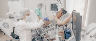
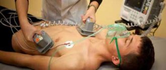
When recording phonocardiography, the patient lies on the couch; the doctor may ask him to hold his breath and take a deep breath. During the examination, the microphone is placed in different areas of the chest in accordance with the auscultatory pattern. Recording can be carried out using additional tests - the introduction of drugs that change cardiac activity or dilate blood vessels.
The duration of the procedure without additional drug load usually does not exceed ten minutes, and the introduction of drugs increases it to half an hour. In most cases, PCG is performed simultaneously with ECG registration (second lead), which allows the sounds to be correlated with the electrical activity of the heart.
Recording sounds
Having learned that auscultation and phonocardiography make it possible to hear various sounds that arise during the work of the heart, let's figure out what exactly they are and what characteristics they have in order to accurately understand the entire need for the procedures:
- The strength of sound is calculated in decibels and represents the sound energy transferred per 1 second per 1 cm2. It is proportional to the amplitude of sound vibrations, so the higher it is, the louder the heartbeats are.
- The frequency of sound is measured in hertz and represents the number of sound vibrations that occur in one second. A person can hear an average sound frequency in the range of 20-20,000 Hz.
- Pure heart sounds on phonocardiography are very rare and represent sound vibrations with one single sound frequency.
- Complex tones are created when a certain number of pure tones are mixed, which happens much more often.
- Noises are multiple sound vibrations that are not connected to each other by the correct ratio, so their presence most often indicates the presence of pathologies.
Basics of FKG
Auscultation provides a large amount of information about the main sounds that appear in the heart during its contraction, but it is to a certain extent subjective, because we all hear differently. In addition, using the ear it is difficult to estimate the amplitude, duration, and interval between individual sound vibrations. For such an analysis, you need a device that impartially records everything that happens in the heart and large vessels.
Cardiac phonocardiography, regardless of its information content, cannot be an independent diagnostic method like ECG or auscultation; it only complements them. It is impossible to decipher its results without detailed knowledge of auscultatory signs of cardiac activity, and for the correct application of sensors, one way or another, you will have to listen to the heart with a phonendoscope.
A little physics
Sound waves characterize vibrations of the walls of the heart, blood vessels, the movement of blood and its impacts on obstacles. The main characteristics of sound are its strength and frequency. Strength is measured in decibels and is proportional to the amplitude of the sound wave. The louder the sound the doctor hears, the greater its strength and greater amplitude will be on the FCG recording.
The frequency of a sound wave is measured in Hertz, which is the number of vibrations of sound per unit time. The human ear hears sounds in the range of 20-20,000 Hz, and everything that lies beyond these limits can only be determined by special equipment. In fact, there is no need to search for sounds that lie beyond the boundaries of our perception, since the heart produces tones in the range of 150-200 Hz, and noises do not exceed 1000 Hz in frequency, so the doctor himself can hear both of them perfectly well. However, in pathology, low-frequency vibrations may occur, which, if audible, are difficult to analyze by the ear, and the third and fourth heart sounds may go unnoticed in the mass of other sound waves, so a detailed characterization and search for such low-frequency waves in heart diseases takes on special meaning.
In cardiology, sounds produced by a beating heart are divided into tones and murmurs. The heart sound is a fairly loud and clear sound. With pathology in the heart, in addition to tones, various types of noise appear. These sounds are not related to each other and have different strengths and frequencies. Both tones and noises in most cases can be detected by auscultation, and at different listening points they will have different volumes.
A phonocardiograph consists of a microphone, analyzing and recording devices. The microphone “hears” heart sounds and noises, the device amplifies them, converts them into electrical signals and records them on paper, similar to what happens with an ECG. The phonocardiograph is equipped with filters to eliminate unnecessary noise and make recording more accurate. The procedure does not cause pain or discomfort and does not require any specific preparation.
Who is assigned to the study?
Now let's find out who is prescribed phonocardiography (PCG) and with what symptoms the patient is sent for such a study. So, this procedure is necessarily prescribed for those who suffer from any defects of the valve apparatus, congenital anomalies of the heart and rheumatism, which is accompanied by inflammation of the organ.
In addition, patients who come to a cardiologist or therapist with complaints of shortness of breath that occurs during physical activity, or pain in the chest and heart, as well as those who have previously suffered a myocardial infarction, have an increased organ size, and those , who had abnormal sounds noted during examination with a stethoscope.
Advantages of FKG
Some people think that if it is possible to undergo auscultation, then cardiac phonocardiography is no longer needed. However, this is not at all true, because this procedure has a number of advantages. And the main thing is that such a study allows us to exclude all subjective factors that may arise as a result of the auscultation technique for recording sounds. Indeed, in this case, it can be affected by the doctor’s hearing, various extraneous noises, device breakdown, and with phonocardiography, all sounds are recorded by a machine, so here the result will be one hundred percent truthful and will allow you to give an accurate assessment of the strength and frequency of the sound, the duration of noises and tones, as well as intervals that occur between them. And if you conduct a FCG and an ECG at the same time, the result of the study will help the doctor see the clearest picture of the patient’s heart condition and prescribe appropriate treatment.
Disadvantages of FKG
However, everything is not so good, because the phonocardiography method has not only advantages, but also significant disadvantages. And the main one is that the phonocardiograph has a very weak sensitivity of the device compared to the human ear, which is why weak sounds that a well-hearing doctor can distinguish during auscultation will not be visible on the phonocardiogram.
In addition, from it it will be impossible to determine the nature of the noise, even subjective, which is completely unique in some cases of heart valve pathologies. And finally, in the case of displacement of the heart or dilatation and hypertrophy of its chambers, it is very difficult to find a place where the phonocardiograph microphone should be placed.
Presence of contraindications
According to cardiologists and therapists, phonocardiography is a procedure that has virtually no contraindications. In addition, this research method is completely safe and painless, so it is prescribed even to those patients who are in serious condition. However, there are still several factors due to which the phonocardiograph microphone cannot be placed close to the chest, and they may become contraindications for this procedure.
Such factors include severe obesity, as well as violation of the integrity of the breast skin such as trauma, burns or wounds. In addition, FCG is not recommended for patients who drank coffee in the morning, as it increases blood pressure, which means the study result will be unreliable.
Research process
A study using the phonocardiography method is a procedure that is carried out using a phonocardiograph, consisting of a microphone and an amplifier, which help to hear all the sounds of the heart, a frequency filter that eliminates interference and makes the sound clearer, and a recording device, thanks to which we get a phonocardiogram in our hands with a detailed description of the strength and frequency of the sound, as well as all murmurs and heart sounds.
To get the correct result, before attaching six microphones to the chest, the doctor listens to the patient, and then lays him horizontally, installs the elements of the device on the chest and examines the work of the heart for ten minutes.
If necessary, the patient may be asked to turn over on his side during the procedure, hold his breath, or take a deep breath. In parallel with the PKG, the patient is additionally given an ECG, which makes it possible to compare all the sounds recorded by the phonocardiograph with the electrical activity of the heart. The entire procedure in this case takes a maximum of 10 minutes.
Advantages and disadvantages of phonocardiography
Like any diagnostic method, FCG has advantages and disadvantages. The advantages are considered:
- The objectivity of the data obtained about the nature of sounds and their graphic display;
- The ability to determine the physical parameters of sound - strength, frequency, interval between tones or noises;
- The ability to objectively compare sounds occurring at a certain interval, which is difficult to do using a phonendoscope;
- Determination of the relationship with the electrical activity of the heart when used simultaneously with an ECG.
Along with the advantages, the FCG method is not without its disadvantages. Of course, the device can “hear” low-frequency vibrations that are inaccessible to the doctor, but still the ear is more sensitive, so some weak sounds in the audibility range can be detected by the doctor, but not displayed on the FCG.
In addition to the low-frequency third, fourth and fifth tones, all other sounds should be audible through the phonendoscope and reflected on the FCG. If the device indicates the presence of sound vibrations that are not audible during auscultation, then the doctor still bases the conclusions on data from listening to the ear (with the exception of low-frequency recordings). Thus, without auscultation, FCG analysis is meaningless.
In addition, the device cannot determine the timbre of the sound; this is done by a person. Timbre is a subjective characteristic, but it significantly helps in diagnosing some valvular heart defects.
To place the microphone at the points where sounds will be heard maximum, preliminary auscultation is used. Many heart diseases are accompanied by changes in its configuration and size, expansion and displacement of the boundaries and, accordingly, the places of best audibility of sound waves, and incorrect position of the microphone will lead to a decrease in sound strength.
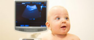
Correct registration and interpretation of PCG data is impossible without previous auscultation, and sounds are assessed based on subjective characteristics determined by the doctor and objective physical data recorded by the device. Recently, PCG has begun to be used in the prenatal diagnosis of fetal hypoxia and congenital heart rhythm disorders.
Study of fetal heart function
Also, using phonocardiography, the physiology of the sound waves emitted by the beating heart of the baby in the womb can be studied to find out how the fetus is developing and whether there are any complications. In this case, the microphone is attached to the area of the pregnant woman's abdomen where the tones of the fetal heart are best heard.
And when the mother begins to give birth, the microphone is moved even lower, to the pubic symphysis. And in this case, FCG is already done from the very contractions until birth, in order to know that they are proceeding normally, focusing on the phonocardiogram indicators, where it should be clear that the heart sounds are even and are heard at equal intervals. The only thing is that when a woman pushes and contractions begin, the amplitude of the second tone increases, but then the height levels out again.
But if phonocardiography of the fetus shows a heart murmur, an increase or decrease in the rhythm frequency, a change in the tone or strength of the sound, or its splitting, this may indicate peritoneal asphyxia, and then the gynecologist must do something to ensure that the birth proceeds normally.
Sound amplitude
The main factor that phonocardiography examines is the amplitude of sounds, an increase or decrease in which indicates the presence of various diseases in the patient.
- The appearance of sounds III and IV on the phonocardiogram may indicate myocardial infarction or hypertension, while the first often signals heart failure, and the second indicates increased contraction of the atria or ventricular hypertrophy.
- An increase in tone II may indicate pulmonary or arterial hypertension, and a decrease in tone may indicate a deterioration in blood flow through the pulmonary artery or aortic valve insufficiency.
- Strengthening of the first tone can be observed with hyperthyroidism, anemia or stenosis of the bicuspid valve, and its weakening indicates dilation of the bronchioles, inflammation of the pericardium or exudative pleurisy of the lung on the left side, in addition, a weak first tone occurs in overweight people.
- An increase in the amplitude of the first tone may indicate mitral stenosis, and its decrease may indicate pathology of the heart muscle, cardiac decompensation, or slowing of atrioventricular conduction.
- A reduction in the intensity of the first tone on the phonocardiogram may be caused by problems with the contractile function of the left ventricle. This phenomenon can often be observed in diseases such as chronic cardiac ischemia, myocardial dystrophy, rheumatic carditis or cardiomyopathy.
Normal FCG
The patient, having received the result of a particular study, usually immediately makes efforts to independently decipher it. If in the case of blood tests this can, at the very least, be done independently, then FCG is a method that lies beyond the capabilities of the average person. To interpret data, you need to master the auscultation method, the basics of which are taught at universities, and experience is gained through practice in the field of cardiology and therapy.
A healthy heart allows you to hear the first and second sounds - these are normal and basic indicators of its functioning.
1,2,3,4 heart sounds on FCG and comparison with ECG
The first sound is formed when the valves located between the atria and ventricles close. The “slamming” of the valve flaps produces a fairly loud sound of medium frequency, the amplitude of which on the FCG reaches 25 mm, and the duration is 0.15 seconds. It is impossible to isolate the sounds of each valve separately; everything happens very quickly and sound vibrations, overlapping each other, give a single first tone.
Correlating the characteristics of the first tone with ECG data, the time of its appearance is given important diagnostic significance. Normally, the interval between the beginning of the Q wave and the beginning of the first tone on the PCG is no more than 0.06 s, and its increase indicates pathology.
The second heart sound characterizes the closure of the pulmonary artery and aortic valves. It is best heard in the second intercostal space to the right and left of the sternum, that is, at the points closest to the base of the heart. Unlike the first tone, the second has two components coming from each valve. These sounds can be distinguished both by the ear and on the device. This phenomenon is especially noticeable in thin people, teenagers and children.
The first sound wave arises from the closure of the aortic valve, it is louder, and its amplitude can exceed the sound from the pulmonary valve by one and a half to two times. This is due to the fact that the pressure in the aorta is much higher than in the pulmonary artery. The next sound appears when the pulmonary valve flaps “slam”, and the interval before its appearance should not exceed 0.06 seconds, otherwise we can talk about the splitting of the second tone. On the ECG, at the moment the second tone appears, the T wave has already ended, or a maximum of 0.4 seconds has passed from the moment of its end.
The third and fourth heart sounds are recorded on FCG quite rarely. When identifying them, you should not make hasty conclusions, and talk about pathology only after a detailed examination. Normally, the third tone can be present in subtle adults, children and adolescents; it consists of sounds produced by the vibration of the walls of the ventricles, so changes in the heart muscle may well be accompanied by its appearance.
It is worth noting that the appearance of the third tone is associated with physical activity, which often occurs in healthy people. It is difficult to listen to it, and the recording occurs in the low-frequency range.
The fourth tone is rarely detected, but, unlike the third, it often indicates pathology. On the ECG it appears after the maximum of the P wave; this noise is low-amplitude and low-frequency.
Video: normal heart sounds, educational video
Heart murmurs
Of no small importance when decoding a phonocardiogram are recorded heart murmurs, which can also be used to judge whether this human organ is functioning well.
- Presystolic murmur may indicate the development of mitral stenosis.
- Oval or diamond-shaped noise on the phonocardiogram indicates the development of aortic stenosis.
- Diastolic murmur may herald the development of aortic insufficiency.
- A band-like systolic murmur that occurs at a low frequency may indicate tricuspid valve insufficiency.
- Functional murmurs occur only in children and can indicate both the development of certain pathologies in them and the health of the child, so they should be judged only in conjunction with all other factors of the PCG and ECG.
CHOU DPO Institute of Resort Medicine and Tourism
INTRODUCTION………………………………………………………………………………..3
- Trends in the development of functional diagnostics in the Russian Federation
Federations………………………………………………………………………………5
- Review of leading methods of functional diagnostics…………………..13
- Possibilities of modern functional diagnostics
and its limitations……………………………………………………………17
CONCLUSION………………………………………………………………………………….19
REFERENCES……………………………………………………….20
Functional diagnostics is a section of diagnostics, the content of which is an objective assessment, detection of deviations and establishment of the degree of dysfunction of various organs and physiological systems of the body based on measuring physical, chemical or other objective indicators of their activity using instrumental or laboratory research methods. The development of functional diagnostics was a direct consequence and practical expression of the physiological direction, which was established in medicine thanks to the achievements of physiology and the works of major clinicians at the turn of the 19th-20th centuries.
It is known that dysfunction of an organ is not always proportional to the amount of structural changes found in it. For example, severe breathing disorders in bronchial asthma or hemodynamics in hypertension are possible with relatively minor morphological changes. While in case of significant structural damage to the organ, for example, when about 2/3 of the pancreas is replaced by a tumor, clinical signs of its functional failure under normal load conditions may be absent.
Meanwhile, limitations in life activity in various diseases are directly related to disorders of the functions of any organs or physiological systems and are proportional to the degree of these disorders. Therefore, along with the morphological, etiological and pathogenetic diagnosis of the disease, the identification and assessment of the degree of impairment of a specific function is the most important part of the diagnosis and is reflected in the formulated clinical diagnosis of the disease. In healthy individuals, the study of the functional reserves of the body, primarily the respiratory and circulatory systems, is carried out for the purpose of predicting and monitoring individual adaptation of a person to extreme environmental conditions (for example, on polar expeditions), sports loads, during professional selection and medical supervision of submariners, divers, pilots, cosmonauts, etc., and in children and adolescents - in order to monitor the correspondence of the development of physiological systems to age.
The purpose of the study is to study modern functional diagnostics in clinical practice, to determine its new capabilities and limitations.
1. TRENDS IN THE DEVELOPMENT OF FUNCTIONAL DIAGNOSTICS
Modern medical diagnostics are used at all stages of the diagnostic and treatment process from diagnosis and throughout the entire treatment phase, and then as a follow-up. Its capabilities are much greater than those of all modern methods of treatment. If quite recently the attending physician at the patient’s bedside could independently make a preliminary diagnosis and prescribe treatment, now a whole team of diagnostic specialists does this together with him.
And to make constructive decisions for the future, you need to look back. First, it should be noted that the specialty “functional diagnostics” exists only in Russia, or more precisely, in the CIS countries, and has no analogues in the healthcare systems of the European Union countries that signed the Bologna Agreement. And this is not a drawback, but rather an achievement of Russian healthcare, which has translated the discoveries of domestic medical science into clinical practice.
The emergence of independent diagnostic specialties in world and domestic medicine is associated with the great scientific discoveries of the 20th century, some of which were awarded Nobel Prizes. The first of them, the discovery of X-rays by Wilhelm Conrad Roentgen (Decision to award the Nobel Prize on November 12, 1901), served as the basis for the emergence of the specialty “medical radiology” and later “radiation diagnostics”. The second is the development of an apparatus for recording electrical signals of the heart by Willem Einthoven, and the method of “electrocardiography” (Decision to award the Nobel Prize on October 23, 1924).
At the beginning of the twentieth century, Russia was the center of the emergence of several world-famous “physiological” schools, which developed a number of important physiological concepts. Among them, the most famous are the schools of Academician I.M. Sechenov (reflex theory), V.M. Bekhterev (fundamentals of psychophysiology), academician I.P. Pavlov (physiology of digestion, physiology of higher nervous activity), academician V.V. Parin (research on reflex regulation of pulmonary circulation, cardiac physiology), academician P.K. Anokhin (theory of functional systems of the body). The results of scientific research by physiologists were quickly introduced into the work of clinical departments of hospitals and other medical institutions in Russia.
For example, the first Russian Nobel laureate I.P. Pavlov personally contributed to the creation of physiological laboratories at clinics, where clinical and physiological seminars, the so-called famous “Pavlovian Wednesdays,” were regularly held. In the depths of scientific schools, and independently, talented scientists and doctors in alliance with engineers developed new technical devices for research.
The appearance in Russia of a device for measuring blood pressure at the beginning of the twentieth century (1905) developed by the Russian military doctor N.S. Korotkov, became a new impetus for the development of diagnostics.
At the same time, new scientific data on the relationship between the nervous and cardiovascular systems have become widespread (K.M. Bykov, V.Ya. Danilevsky, L.A. Orbeli, V.N. Chernigovsky, N.N. Anichkov, etc.) .
In large surgical clinics under the leadership of famous Russian academicians such as A.N. Bakulev, B.V. Petrovsky, A.A. Vishnevsky, N.N. Burdenko and others, the first specialized laboratories of clinical physiology arose. They were essentially intended to introduce the achievements of experimental science into practice. Here new diagnostic devices appeared, algorithms were developed for conducting research by doctors - “clinical diagnosticians”. In the depths of large scientific surgical centers, new diagnostic information, which was previously obtained invasively during operations, was of particular value.
In therapeutic clinics, the emergence of such diagnostic departments meant the possibility of monitoring the patient’s condition throughout the entire period of treatment, and after discharge from the hospital.
The first functional diagnostics room was organized on the initiative of the head of the department of therapy of the Central Institute for Advanced Medical Studies, Professor D.D. Pletneva. In 1930, on the basis of the MONIKI therapeutic clinic, the first functional diagnostic department was created, and later, in 1932, the first research institute was organized - “Research Institute of Functional Diagnostics and Experimental Therapy”. The first diagnostic rooms, having appeared in the institute's therapeutic clinic, then began to appear everywhere, first in hospitals, and then at the outpatient level, thereby solving the problem of rapid primary diagnosis, primarily of cardiovascular pathology.
President of the USSR Academy of Medical Sciences A.A. In 1945, Bogomolets, at one of the first post-war plenums of the Academy, announced the emergence of a new specialty in Soviet medicine, called “clinical physiology.”
The specialty has firmly occupied its own special niche among other clinical specialties; its “legal successor” has become “functional diagnostics”.
New scientific schools using hardware technologies, primarily in cardiology (A.F. Samoilov, P.E. Lukomsky, A.L. Myasnikov, etc.) contributed to the further development of instrumental diagnostics in the clinic and the emergence of specialized specialists in this field. So, the specialty “Functional Diagnostics”, having arisen as a result of the introduction of the achievements of Russian scientific schools into the clinic, continues its development even now.
The first regulatory document “Regulations on the Electrocardiographic Room”, approved on April 21, 1954 by the Main Directorate of Medical and Preventive Care of the USSR Ministry of Health, marked the beginning of a new stage - the introduction of the first hardware ECG method into widespread medical practice.
At the same time, other methods for studying the heart and blood vessels appeared. All of them were introduced into the practice of functional diagnostic rooms.
The second half of the 20th century was marked by another important event - the advent of ultrasonic devices.
Ultrasound examination of “moving structures” of the heart and blood vessels has become an important addition to existing traditional methods of functional diagnostics, such as ECG, PCG, RVG, etc.
Later, the ultrasonic method replaced some of them. The development of ultrasound methods and the widespread introduction of computer technologies have changed the capabilities and worldview of a functional diagnostics doctor.
Today, the study of the function of the cardiovascular system is carried out with the mandatory use of a complex of functional methods, including ultrasound.
Today, functional diagnostic departments use devices of both domestic and imported origin. During the period of modernization of domestic healthcare and updating of medical equipment, it is appropriate to recall another fact of our history. In the USSR in 1967, the production of domestic medical equipment and instruments was separated into a separate industry. Equipping hospitals, clinics, clinics, and research institutions with sophisticated medical equipment required the involvement of many ministries and departments in this matter.
This was recorded in a number of resolutions of the CPSU Central Committee and the USSR Council of Ministers in 1977-1980. On the initiative of the USSR Minister of Health, Academician B.V. Petrovsky carried out a large amount of work on the creation of domestic medical equipment and instruments, many types of which are still not inferior to the best foreign models. The creation of a special government commission in the USSR, which obliged industrial ministries to develop the necessary apparatus and instruments for the needs of the Ministry of Health, became important for healthcare in general. In accordance with the decree, the ministries were assigned areas of development, which ensured their specialization and coordination of research and development work.
The emergence of a new diagnostic specialty, “functional diagnostics” (FD), was vitally important for the practicing physician. But if its emergence was associated with the introduction into the clinic of the results of research from scientific schools and the emergence of various diagnostic devices, today the opposite situation has arisen, when there is a medical specialty, but no scientific one.
The lack of a scientific specialty “functional diagnostics” deprives the possibility of further harmonious development of all instrumental diagnostic areas.
Without the possibility of scientific comprehension and analysis, without the formulation of diagnostic research concepts in the form of master's and doctoral dissertations, it is impossible to talk about the future of any direction in medicine. However, there is one convincing fact that this problem will be solved. This fact is the formation in 1996 of the Russian Association of Functional Diagnostics Specialists (RASD). By this time, in the depths of the scientific and medical community of PD specialists, the idea of uniting into a public organization was born in order to solve current theoretical, scientific and practical problems, determine priority directions for the development of PD, and protect the rights and professional interests of members of the Association.
An initiative group of specialists from scientific organizations that include large PD departments: MONIKI, RMAPO, MMA named after. Sechenov, RKNPK (Cardiocenter) - the draft Charter of “RASFD” was prepared, and the first documents were prepared for registration of a new public organization.
At the same time, the quarterly peer-reviewed journal of the RASFD “Functional Diagnostics” was established. The chief freelance specialist of the Ministry of Health of the Russian Federation, head of the department of functional diagnostics of MONIKI, Professor Sergei Sergeevich Koltsun, was elected the first President of the Association.
Thus, the history of the development of our specialty is twice connected with the Moscow Research Institute of Clinical Medicine named after. F.M. Vladimirsky, where for the first time in 1932 the Research Institute of Functional Diagnostics and Experimental Therapy was organized (under the leadership of Professor D.D. Pletnev).
The first composition of the RASFD Council included: Beresten N.F., Dyachenko T.Yu., Ivanov G.G., Kechker M.I., Makareva N.M., Rogoza A.N., Ryabykina G.V., Sakhno Yu.F., Fedorova S.I. The audit commission included: Pronina V.P., Massarygin V.V., Laskarzhevskaya M.A.
Today, not only doctors, but also leading specialists of Russian scientific organizations are concerned about the current situation. RASFD, at each conference since 1997, has included in the resolution a decision on the need to recreate the scientific specialty “Functional Diagnostics”. It is necessary to include the specialty “Functional Diagnostics” in the list of scientific medical clinical specialties (Order of the Ministry of Education of the Russian Federation of February 25, 2009 No. 59 “On approval of the nomenclature of specialties of scientific workers”).
The scientific specialty “functional diagnostics” should contribute to the study of the functional state of organs and systems of the human body in normal conditions and in pathology using various instrumental methods. This step will lead to an influx of new personnel into the specialty, and new opportunities will appear in mastering and studying the mechanisms of formation of pathological processes studied using various technologies.
This decision will also lead to the re-creation at a qualitatively new level of what was once organized by Professor D.D. Pletnev – Research Institute of Functional Diagnostics and Experimental Medicine.
This step is important for all departments of functional diagnostics of medical universities that do not have clear guidelines for training highly qualified PD specialists for practical medicine.
The importance of developing functional diagnostics as a scientific specialty is also confirmed in the Project “Strategies for the development of medical science in the Russian Federation for the period until 2025”, developed by the Ministry of Health of the Russian Federation, the Russian Academy of Medical Sciences, the Russian Academy of Sciences, Moscow State University. M.V. Lomonosov in accordance with Decree of the President of the Russian Federation of May 7, 2012 No. 598 “On improving state policy in the field of healthcare.”
The document on scientific directions of functional diagnostics states that “a new generation of functional diagnostic methods will be created based on multifunctional monitoring of patients in real life conditions; new stress tests to detect coronary heart disease using new technologies for obtaining and analyzing the electrical field of the heart (microalternation PQRST and other existing or new ones); complex diagnostic systems using methods for studying the electrical field of the heart in combination with various imaging methods and constructing corresponding electromechanical models of the myocardium.
The creation of new methods for identifying preclinical damage to target organs and prognostically unfavorable dysregulatory changes in the cardiovascular system, adapted to the requirements of preventive medicine, and their widespread implementation in practice will make it possible to diagnose cardiovascular diseases in the early stages...”
2. REVIEW OF LEADING METHODS
Functional diagnostics currently serves as a good aid for the clinician in making a diagnosis. The equipment and methods themselves are being improved every year, and the field for diagnostics is expanding. The clinician can study the functioning of the organ and obtain more information than is available to the senses: the eyes when examining a patient, the ear when performing auscultation, etc.
The study can be carried out on healthy individuals or to diagnose certain diseases. In healthy individuals, functional tests are carried out to assess the function of organs or systems before complex expeditions, business trips, or work that requires great physical and mental effort. To diagnose diseases, a specialist is referred for a specific test.
Method for recording electrical potentials of the heart. Einthoven was the first to conduct experiments on recording electrical impulses of the heart. In 1924, he was awarded the Nobel Prize in Medicine or Physiology for the creation of the first ECG machine.
Typically, the ECG is recorded in 12 leads: 3 standard leads, 3 enhanced leads and 6 chest leads. Additional leads are used to diagnose myocardial infarction of the posterobasal sections of the left ventricle (unipolar or bipolar according to the Sky). The recording is made in a calm state of the patient with normal breathing. First, standard leads are recorded on film, then reinforced leads and chest leads.
Holter monitoring ECG / HM-ECG / “Holter” - the method allows you to record ECG potentials throughout the day. The subject keeps a diary in which he records episodes of physical activity, emotional stress, anxiety, and food intake. Marks the time at which this event occurred. The patient’s complaints (pain in the heart area, interruptions in heart function, rapid heartbeat, etc.) are also recorded in the diary. The recording is then transcribed using a computer program, and the results are compared with the entries in the diary. Based on the data obtained, the doctor makes the correct diagnosis.
The principle is the same as the “holter”. During the day, the patient wears a device that measures his blood pressure at specified intervals. Using the same principle, the patient keeps a diary. There are some additional rules. While measuring blood pressure, the patient's hand should remain motionless. For example, if the device starts to work while walking, you need to stop and extend your arm along your body. When measuring blood pressure at rest, you must follow all the rules that are established for measuring pressure with a conventional tonometer. Indications: “white coat” hypertension, hypertension in combination with other heart diseases, symptomatic hypertension, diagnosis of hypotensive conditions, correction of drug treatment methods. In the outpatient department, devices are used for non-invasive measurement of blood pressure using the auscultatory or oscillometric method.
An invasive method of measuring blood pressure is used in cardiac surgery.
For diagnosis, the average result of systolic, diastolic and pulse blood pressure, pressure variability and frequency of increase above the figures of 140 and 80 mmHg are important. or decrease to 90 and 60 mmHg or less.
Electroencephalography is a method for studying the biopotentials of the brain. EEG is recorded using electrodes placed on the head. There are two ways of recording EEG - monopolar and bipolar. The bipolar method makes it possible to detect pathological formations in the brain (for example, tumors) by recording the potential that arises between two active electrodes. The monopolar method of EEG recording makes it possible to study oscillations and their amplitude, assess brain activity, and diagnose dystrophic and degenerative diseases of the brain.
Electroneuromyography
The method allows you to study the function of excitable tissues, which include nervous and muscle tissue. The condition of the entire path that the nerve impulse travels is assessed: starting from the roots of the spinal cord and ending with the neuromuscular synapse. This technique allows you to distinguish between damage to a muscle, neuron or pathway. It may be useful in diagnosing demyelinating diseases.
Spirography is a method for studying the function of external respiration. Tidal volume, forced expiratory rate, residual air volume after maximum exhalation and other indicators are measured. In addition, you can conduct a test with bronchodilators and evaluate how the spirogram readings will change.
The test is valuable in diagnosing bronchial asthma and chronic obstructive pulmonary diseases.
3. POSSIBILITIES OF MODERN FUNCTIONAL
Currently, new computer electrocardiographs are being installed in hospitals in the department of functional diagnostics. Modern equipment is designed to conduct ECG studies. Unlike a classic electrocardiograph, which produces the examination result as a printout on thermal paper, a computer electrocardiograph transmits the patient’s electrocardiogram to a computer via a local network in the department of functional diagnostics and stores it digitally in a database. To analyze the ECG, the department has connected workstations of doctors, who make a conclusion within a few minutes.
A computer electrocardiograph is a complex consisting of an ECG module, a computer with software for recording, analyzing, storing and transmitting an ECG.
Previously, to decipher the electrocardiogram, it was necessary to deliver it to the doctor, which took time. Now ECG studies of patients from hospital departments and clinics will be sent to specialists of the functional diagnostics department in real time.
Until recently, the result of an ECG examination was pasted into the patient’s chart. It took time to find the necessary entries in the map and compare them with each other. In addition, thermal paper printouts faded over time and became unreadable. With the advent of computer technology, research results are in electronic form, which eliminates the storage of an archive of paper films. New electrocardiograms can be printed on regular office paper.
Also among the advantages of modern equipment that is operating in the hospital today in test mode is ECG visualization. When a doctor writes a conclusion not based on a printout by visual analysis, but has the opportunity to enlarge the image of the electrocardiogram on the monitor screen and display several ECGs for comparison.
CONCLUSION
A functional diagnostics doctor provides qualified specialized care in his area of expertise. He conducts research into the function of a particular system of the human body using special equipment to determine the state of organs and tissues, determine pathological conditions in the early stages of development, and their characteristics.
The doctor examines patients before surgery, during certain stages of medical examination, pregnant women during routine examinations, etc. Large hospitals have functional diagnostic departments. In smaller ones there is a functional diagnostics room.
The study of the functional ability of a particular organ is important when examining both an adult patient and a child. Functional methods allow you to answer a number of questions. Firstly, the results of the study are used to judge how the disease affects the function of an organ or system. Secondly, based on certain parameters, we can judge the compensatory capabilities of the body. Thirdly, by testing with medications or some other influences (physiotherapy, physical activity, etc.), we help the patient choose an adequate treatment and regimen. And finally, by conducting dynamic monitoring of the patient with repeated functional examination, the doctor can judge the effectiveness of treatment, carry out timely correction of treatment and rehabilitation measures, and often predict the course of the disease.
- Kondrashin A.V., Karasev V.S. Functional diagnostics of an adaptive control system: problem statement and its solution // Izvestia YuVU. – 2021. – No. 8 (181). – pp. 79-88.
- Prokofiev A.B., Rudnev S.G., Marinin V.F., Kukes I.V. The importance of modern methods of functional diagnostics in clinical studies of drugs used in cardiological practice // Bulletin of the Scientific Center for Expertise of Medical Products. – 2014. – No. 3. – P. 19-23.
- Sokolov A.A. Echocardiography and functional diagnostics // Siberian Medical Journal (Tomsk). – 2021. – T. 22. – No. 3. – P. 54-58.
- Functional diagnostics: textbook / comp. Ozhev B.V. – Maykop: MSTU Publishing House, 2015. – 64 p.