Cardiac axis and ECG
Deviation of the electrical axis of the heart to the left
The human heart has the ability to contract. Electrical impulses sequentially cover the chambers of the heart, originating in the atrial sinus node. If you imagine the course of these pulses in the form of directed vectors, you will notice that they have a similar direction. By summing the directions of the vectors, one main vector can be obtained. This will be the electrical axis of the heart (EOS).
Functional diagnostic doctors often determine EOS from a cardiogram visually, but it is more accurate to do this using special tables. If you look carefully at the QRS complex in leads I, II, III on the ECG, you can see that R II>RI>RIII, this means that the EOS on the cardiogram is normal.
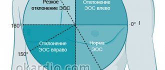
If it is difficult for a doctor to visually determine the axis of the heart, he determines the alpha angle and calculates the EOS using special tables. Without delving into the course of measurements, we note that for a normal EOS angle alpha (RII>RIII), then the doctor’s conclusion will be as follows: deviation of the electrical axis of the heart to the left. EOS deviation is confirmed when the alpha angle is in the range from 00 to -900.
The influence of the anatomical position of the heart on the electrical axis of the QRS complex
Confirmed by the effect of breathing . When a person inhales, the diaphragm lowers and the heart takes a more vertical position in the chest, which is usually accompanied by a vertical displacement of the EOS (to the right). In patients with pulmonary emphysema, an anatomically vertical position of the heart and an electrically vertical mean electrical axis of the QRS complex are usually observed. On the contrary, when exhaling, the diaphragm rises and the heart takes a more horizontal position in the chest, which is usually accompanied by a horizontal displacement of the EOS (to the left).
When does the heart axis “go to the left”?
Sharp deviation of the electrical axis of the heart to the left
The conclusions of a functional diagnostics doctor about the deviation of the cardiac axis to the left are not an independent diagnosis. But they always give reason to wonder why the heart axis “went to the left.” A slight shift of the EOS to -190, as well as its semi-vertical position, in some cases is not considered a pathology. This position of the axis can be observed in healthy, tall, thin people, in athletes with a trained heart, in children with an asthenic physique, and with a high position of the diaphragm dome.
If the cardiac axis is significantly deviated to the left, then this pathological condition indicates problems with the heart; the cause of such a displacement must be established. After all, this symptom can sometimes be the first “bell” in case of pathology of the heart and blood vessels. According to some data, a deviation of the electrical axis of the heart to the left up to -29-300 is sometimes called a slight deviation, and if the angle is from -450 to -900 they speak of a sharp deviation.
The location of the electrical axis is normal
In healthy people, the electrical axis of the heart coincides with the anatomical axis of this organ. The heart is located semi-vertically - its lower end is directed down and to the left. And the electrical axis, like the anatomical one, is in a semi-vertical position and tends down and to the left.
The standard alpha angle is from 0 to +90 degrees.
Norm of angle alpha EOS
The location of the anatomical and electrical axes depends to some extent on body type. In asthenics (thin people with tall stature and long limbs), the heart (and, accordingly, its axes) is located more vertically, while in hypersthenics (short people with a stocky build) it is more horizontal.
Normal alpha angle depending on body type:
| Body type | Minimum | Maximum |
| Normosthenics | +30 | +70 |
| Asthenics | +70 | +90 |
| Hypersthenics | 0 | +30 |
A significant displacement of the electrical axis to the left or right is a sign of pathologies of the conduction system of the heart or other diseases.
A deviation to the left is indicated by a minus alpha angle: from -90 to 0 degrees. About its deviation to the right - values from +90 to +180 degrees.
However, it is not at all necessary to know these numbers, since in case of violations in the ECG interpretation you can find the phrase “EOS is deviated to the left (or right).”
Pathological causes of EOS shift to the left
Pathological conditions in which there is a displacement of the cardiac axis to the left
As mentioned above, a slight deviation of the EOS to the left can be considered by doctors as a variant of the norm, if, after a more thorough examination, the doctor did not identify any diseases in the patient and the patient’s health is good. If the EOS is significantly deviated to the left, or the patient has health problems due to minor ECG changes, the following pathological conditions should be suspected, in which a shift to the left of the cardiac axis is most common:
Left ventricular hypertrophy
The deviation of the cardiac axis to the left with an enlargement of the left ventricle is quite understandable, because physiologically this chamber of the heart is already the most powerful in terms of mass. This means that the vector of the heart will “take over” the left ventricle. And the more it increases in size and grows, the more the EOS will “move to the left.” This pathology occurs with high blood pressure or arterial hypertension, when the chambers of the heart, unable to withstand the increased pressure and load, begin to compensatory gain weight - hypertrophy. Hypertrophy as one of the symptoms occurs in heart failure, atherosclerotic vascular changes, angina pectoris, cardiac asthma, and cardiomyopathies.
Conduction disorders
Left bundle branch block
Disturbances in the conduction system will lead to changes in the cardiac vector and deviation of the cardiac axis. This is most often observed with blockade of the left bundle branch, or with blockade of its anterior superior branch. There are other ECG signs that can help diagnose this type of arrhythmia. Holter ECG monitoring will also help in establishing the diagnosis.
Special forms of ventricular tachycardia
Some forms of ventricular tachycardia may also be the reason why EOS values are far from normal.
Heart defects
Heart defects, the ECG symptom of which can be the axis of the heart moving to the left, by their nature can be either congenital or acquired. Defects of any etiology, accompanied by overload of the left heart chambers, will be characterized by this ECG symptomatology.
Based on the reasons described above for EOS deviation, we can conclude that a shift to the left of the cardiac axis is not such a harmless ECG sign. It may indicate the presence of quite serious problems in the patient’s body. But at the same time, don't panic! If the patient is in good health, has a stable ECG for several years, and in the absence of confirmatory data on pathological changes in the heart and blood vessels after a thorough examination, a slight deviation of the cardiac axis to the left may be a variant of the norm! But the conclusion that this is the norm can be made by a doctor after a thorough examination of the patient, and in the absence of data on the pathology of the cardiovascular system. What examinations should a doctor prescribe when diagnosing a patient with a shift to the left of the heart axis?
Additional diagnostics
To find out the reasons for the EOS deviation, the ECG is analyzed in detail. They may also assign:
- EchoCG (ultrasound of the heart) - to identify possible organ defects.
- Stress echocardiography – ultrasound of the heart under stress – for diagnosing ischemia.
- Angiography of the coronary vessels - their examination to identify blood clots and atherosclerotic plaques.
- Holter monitoring – recording an ECG using a portable device throughout the day.
After a detailed examination, appropriate therapy is prescribed.
A set of examinations to clarify the diagnosis
Taking a repeat cardiogram
- Repeated ECG. It is imperative to take a repeat cardiogram, especially if EOS displacement is detected for the first time and previous ECGs were normal. An error in the application of electrodes, which may show a distorted result, or a malfunction of the cardiograph cannot be ruled out. It is also always necessary, if possible, to compare a “fresh” ECG with a previous cardiogram to assess the dynamics of the patient’s condition and monitor changes in the work of the heart.
- Ultrasound of the heart. The most informative way to tell about the condition of the heart, its chambers, cardiac ejection fraction, and the flow of blood through the cardiac cavities is with ultrasound or echocardiography. This examination method can be supplemented, if necessary, with Doppler sonography.
- Holter ECG. If the doctor suspects a patient has conduction disturbances or rhythm disturbances, then Holter ECG monitoring will be a reliable assistant in making the diagnosis. A daily recording of a cardiogram will allow the doctor to “catch” the arrhythmia and see in which part of the heart the conductivity is changed. To ensure that Holter data is not distorted, the patient should be given detailed instructions on how to behave during the study.
24-hour blood pressure monitoring - ABPM. With high blood pressure in combination with LV hypertrophy and cardiac axis deviation, the doctor may recommend 24-hour blood pressure monitoring to the patient. This diagnostic method helps to establish a more accurate degree of hypertension and help in selecting optimal doses of antihypertensive drugs.
- Consultation with a cardiac surgeon. Consultation is required if heart defects are suspected, or after they are detected by ultrasound.
It should be understood that deviation to the left of the EOS is not a diagnosis, but an ECG sign, which can be either a variant of the norm or a symptom of numerous diseases. Only a doctor can make a conclusion about what information this symptom carries, after carrying out a set of diagnostic procedures.
Is it necessary to treat an axle tilted to the left?
Is treatment necessary?
As the only isolated ECG sign - no. If this symptom is one of the others in the presence of a disease in the human body, the disease certainly needs to be treated. Treatment tactics depend directly on the disease that caused changes in the direction of the cardiac axis. In case of hypertension, which has led to an enlargement of the left ventricle, an adequate selection of antihypertensive drugs is necessary. For arrhythmias - antiarrhythmic drugs, or, if indicated, implantation of an artificial pacemaker. For diagnosed heart defects, surgical treatment is performed according to indications.