8379
This term originates from Latin and means “closure.”
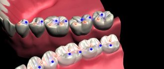
Central occlusion is a state of evenly distributed tension of the jaw muscles, while ensuring simultaneous contact of all surfaces of the elements of the dentition.
The need to determine central occlusion is to correctly manufacture a partial or removable denture.
Main features
Experts have determined the following indicators of central occlusion:
- Muscular. Synchronous, normal contraction of the muscles responsible for the functioning of the lower jaw bone.
- Articular. The surfaces of the articular heads of the lower jaw are located directly at the bases of the slopes of the articular tubercles, in the depths of the articular fossa.
- Dental:
- full surface contact;
- opposite rows are brought together so that each unit is in contact with the same and the next element;
- the direction of the upper frontal incisors and the similar direction of the lower ones lie in a single sagittal plane;
- the overlap of the elements of the upper row of fragments of the lower one in the front part is 30% of the length;
- the anterior units contact in such a way that the edges of the lower fragments abut the palatine tubercles of the upper ones;
- the upper molar comes into contact with the lower one so that two-thirds of its area is combined with the first, and the rest with the second;
If we consider the transverse direction of the rows, then their buccal tubercles overlap, while the tubercles on the palate are oriented longitudinally, in the fissure between the buccal and lingual of the lower row.
Signs of correct row contact
Are common:
- the rows converge in a single vertical plane;
- incisors and molars of both rows have a pair of antagonists;
- there is contact between units of the same name;
- the lower incisors do not have antagonists in the central part;
- the upper eighths have no antagonists.
Applies to anterior units only:
- if we conditionally divide the patient’s face into two symmetrical parts, then the line of symmetry should pass between the front elements of both rows;
- the upper row of fragments overlaps the lower one in the anterior zone to a height of 30% of the total crown size;
- the cutting edges of the lower units are in contact with the tubercles of the inner part of the upper ones.
Applies only to lateral ones:
- the buccal distal cusp of the upper row is based in the space between the 6th and 7th molars of the lower row;
- the lateral elements of the upper row close with the lower ones in such a way that they fall strictly into the intertubercular grooves.
Let's find out together how dental impressions are made and what modern material is used.
Read here about the purpose of filing the front teeth.
At this address https://orto-info.ru/ortodonticheskoe-lechenie/podgotovitelnyiy-period/separatsiya-zubov.html we will talk about the consequences of teeth separation.
Diagnosis of mesial occlusion
As can be seen from the above, mesial occlusion has signs. And there are reasons. And these concepts should not be confused. Those. You should not treat the signs (“third class closure” or “protruding lower jaw”). The cause needs to be treated. Before that, of course, it was determined diagnostically.
Diagnosis is the first and key step in the treatment of mesial occlusion. Do you want to know why it is impossible without diagnosis?
In addition, the causes of mesial occlusion, as can be seen from the text above, can be divided into dental and skeletal. And skeletal causes can be divided into jaw and postural (related to posture). And all these reasons can be combined in various ways.
In general, in order to clearly understand the cause of mesial bite, in each specific case, a diagnosis is needed. Only after analyzing the diagnostic data will it become clear how and with what to treat mesial occlusion in this particular patient.
The main types of diagnostic studies for mesial occlusion are:
- analysis of lateral TRG (teleradiogram). It allows you to identify jaw factors (size and position of the jaws) of mesial occlusion
- analysis of jaw models (dentition). It allows you to identify the “dental” causes of mesial occlusion
Analysis of lateral TRG.
Analysis of jaw models.
Methods used
Central occlusion is determined at the stage of manufacturing prosthetic structures when several units are lost.
This is necessary to ensure the normal functioning of the product and eliminate the occurrence of deviations and problems in the functioning of the temporomandibular joints.
In this case, the height of the lower third of the face is of great importance. However, in the absence of a large number of units, this indicator may be violated and must be restored.
If the patient has partial adentia, several options for determining the indicator are used.
The presence of antagonists on both sides
The method is used when antagonists are present in all functional areas of the jaws.
In the presence of a large number of antagonists, the height of the lower third of the face is maintained and fixed.
The occlusion index is determined based on as many contact zones as possible of the same units of the upper and lower rows.
This option is the simplest, as it does not require the additional use of occlusal ridges or specialized orthopedic templates.
Presence of three occlusion points between antagonists
This method is used if the patient still has antagonists in the three main contact zones of the rows. At the same time, the small number of antagonists does not allow normal positioning of plaster casts of the jaw in the articulator.
In this case, the natural height of the lower third of the face is disrupted, and occlusal ridges made of wax or thermoplastic polymer are used to correctly match the casts.
The roller is placed on the bottom row, after which the patient brings his jaw together. After the roller is removed from the oral cavity, imprints of the antagonist contact zones remain on it.
These prints are subsequently used by technicians in the laboratory to position the casts and create a fully functional and correct, from an orthopedic point of view, prosthesis.
Absence of antagonistic pairs
The most labor-intensive scenario is the complete absence of the same elements on both jaws.
In this situation, instead of the position of central occlusion, the central relationship of the jaws is determined .
The procedure includes the following steps:
- Work on the formation of a prosthetic plane , which is positioned along the chewing surfaces of the lateral units and is parallel to the beam. It is built from the lower point of the nasal septum to the upper edges of the ear canals.
- Determination of the normal height of the lower third of the face.
- Fixation of the mesiodistal relationship of the upper and lower jaws using wax or polymer bases with occlusal ridges.
Checking central occlusion with existing pairs of elements of the same name is carried out by closing the teeth and is carried out as follows:
- a thin strip of wax is placed on the already prepared and fitted contact surface of the occlusal roller and glued;
- the resulting structure is heated until the wax softens;
- heated templates are placed in the patient’s oral cavity;
- After bringing the jaws together, the teeth leave imprints on the wax strip.
It is these fingerprints that are used in the process of modeling central occlusion in the laboratory.
If, during the process of determining occlusion, the surfaces of the upper and lower rollers close, the specialist adjusts their contact surfaces.
Wedge-shaped cuts are made on the upper one, and a certain amount of material is cut off from the lower one, after which a wax strip is glued to the treated surface. After the rows are brought together again, the strip material is pressed into the cutouts.
The products are removed from the patient’s mouth and sent to the laboratory for subsequent production of a prosthesis.
Symptoms
Against the background of damage to the brachiocephalic vessels, there is a decrease in performance, weakness and dizziness . The brachiocephalic trunk is responsible for the blood supply to the soft tissues of the head and the brain. If the left artery is additionally involved in the pathological process, then the clinical picture worsens significantly. Main manifestations:
- pale skin;
- nausea;
- headache;
- pain during physical activity;
- confusion;
- paraplegia;
- swelling and development of necrosis;
- burning sensation or numbness;
- deterioration of visual perception;
- hallucinations;
- difficulty breathing, swallowing;
- speech disorders;
- cardiopalmus;
- absence of pulse in the affected area.
If any of the above symptoms appear, it is necessary to conduct a thorough analysis and diagnosis to identify the true cause and subsequently prevent the development of severe complications.
Calculations for orthopedic purposes
In the process of creating prosthetic structures for malocclusion, an orthopedic specialist takes measurements of the heights of the lower third of the patient’s face using an anatomical and physiological method.
To do this, the height of the bite is measured in a state of complete reduction of the jaws, with central occlusion and in a state of physiological rest.
Payment procedure:
- At the lowest point of the nose , at the level of the nasal septum, the first mark is placed strictly in the center. In some cases, the specialist places a mark on the tip of the patient's nose.
- in the center of the chin , in its lower zone.
- Between the applied marks, the height is measured in the state of central occlusion of the jaws. To do this, bases with bite ridges are placed in the patient’s oral cavity.
- Repeated measurements are taken between marks , but in a state of physiological rest of the lower jaw. To do this, the specialist must distract the patient so that he really relaxes. In some cases, the patient is offered a glass of water. After a few sips, the muscles of the lower jaw really relax.
- The results are recorded. However, the standardized indicator of normal bite height, which is 2-3 mm, is subtracted from the height at rest. And if after this the indicators are equal, we can talk about normal bite height.
If, when measuring height, the calculation results yield a negative result, the lower third of the patient’s face is underestimated . Accordingly, if the result deviates in a positive direction, the bite is overestimated .
What is the significance of teleradiography in orthodontics, and what is revealed with its help.
In this publication we will talk about diagnostic methods in orthodontics.
Here https://orto-info.ru/ortodonticheskoe-lechenie/podgotovitelnyiy-period/miogimnastika.html you are offered myogymnastics exercises used in orthodontics.
List of sources
- Matyushenko A.A. “Pulmonary embolism as a general medical problem”, article in the journal RMZh No. 13 dated 07/03/1999
- Atayan A.A., Kosenkov A.N., Kuznetsov M.R., Chernookov A.I., Ivanova M.I., Khachatryan E.O. “Hybrid tactics in the treatment of acute disorders of mesenteric circulation”, RMZh No. 8(II) dated 10.25.2019
- Yavelov I.S. “The use of anticoagulants during thrombolytic therapy in patients with signs of acute occlusion of the coronary artery: how to individualize treatment?”, RMZh No. 26 of November 30, 2011
Techniques for correct positioning of the lower jaw
Correct positioning of the patient's jaw in the position of central occlusion involves the use of two methods of placement: functional and instrumental.
The main condition for correct placement is muscle relaxation of the jaw muscles.
Functional
The procedure for carrying out this method is as follows:
- the patient moves his head back slightly until the neck muscles tense, which prevents protrusion of the jaw;
- touches the tongue to the back of the palate, as close to the throat as possible;
- at this time, the specialist places his index fingers on the patient’s teeth, lightly pressing on them and at the same time slightly moving the corners of the mouth in different directions;
- the patient imitates swallowing food, which in almost 100% of cases leads to muscle relaxation and prevents jaw protrusion;
- When bringing the jaws together, the specialist touches the surfaces of the teeth and holds the corners of the mouth until it is completely closed.
In some cases, the procedure is repeated several times until complete muscle relaxation and correct reduction of both rows are achieved.
Instrumental
It is performed using specialized devices that copy jaw movements. It is used only in extremely serious situations, when bite deviations are significant and it is necessary to correct the position of the jaw using the physical efforts of a specialist.
Most often, when carrying out this method, the Larin apparatus and special orthopedic rulers are used, which make it possible to record jaw movements in several planes.
Formation of the prosthetic plane
“To draw a plane you need three points”
© Geometry
Occlusal plane
- a plane that passes through:
1) the point between the lower central incisors
2) and 3) points on the external posterior tubercles of the second chewing teeth.
Three points: 1) Between the central incisors 2) and 3) Posterior buccal cusp of the second molar
If you have teeth, then there is an occlusal plane. If there are no teeth, then there is no plane. The dentist's task is to restore it. And restore correctly.
Prosthetic plane
Like the occlusal plane, only on a denture
- this is the occlusal plane of a complete removable denture. It should run exactly where the occlusal plane once was. But the dentist is not a psychic; he cannot see the past. How will he determine where she had a patient 20 years ago?
After many studies, scientists have established that the occlusal plane in the anterior jaw is parallel to the line connecting the pupils. And in the lateral section (this was discovered by Camper) - a line connecting the lower edge of the nasal septum (subnosal) with the middle of the tragus of the ear. This line is called the Camper horizontal.
The doctor’s task is to ensure that the prosthetic plane - the plane of the wax ridge on the upper jaw - is parallel to these two lines (Kamper’s horizontal and the pupillary line).
The doctor divides the entire prosthetic plane into three segments: one frontal and two lateral. He starts from the frontal section. And makes the plane of the frontal ridge parallel to the pupillary line. To achieve this he uses two rulers. The doctor places one ruler at the level of the pupils, and attaches the second to the wax roller.
One ruler is installed along the pupillary line, the second is glued to the bite block
He achieves parallelism between the two rulers. The dentist adds or cuts wax from the roller, focusing on the upper lip. As we described above, the edge of the roller should evenly protrude from under the lip by 1-2 mm.
Next, the doctor forms the lateral sections. To do this, the ruler is installed along the Camper (nose-ear) line. And they achieve parallelism with the prosthetic plane. The doctor builds up or removes wax in the same way as he did in the anterior section.
The ruler along the Camper horizontal is parallel to the occlusal plane in the lateral section
After this, he smoothes the entire prosthetic plane. It is convenient to use for this
Naisha apparatus.
The Naisha apparatus is a heated inclined plane with a wax collector.
The base with bite rollers is applied to the heated surface. The wax melts evenly over the entire surface of the roller, in one plane. As a result, it turns out perfectly smooth.
The melted wax is collected in a wax collector, which is shaped like a blank for new rollers.
 Next, the doctor proceeds to determine the height of the lower part of the face. And again a little theory.
Errors allowed
Creating a prosthetic structure in conditions of malocclusion is a most complex orthopedic procedure, the quality of which depends 100% on the qualifications of the specialist and a responsible approach to work.
Violations in determining the position of central occlusion can lead to the following problems:
The bite is too high
- The folds of the face are smoothed, the relief of the nasolabial zone is poorly defined;
- the patient's face looks surprised;
- the patient feels tension when closing the mouth, while closing the lips;
- the patient feels that during communication the teeth are knocking against each other.
Low bite
- The folds of the face are strongly pronounced, especially in the chin area;
- the lower third of the face visually becomes smaller;
- the patient becomes like an elderly person;
- the corners of the mouth are lowered;
- lips sink;
- uncontrolled salivation.
Permanent anterior occlusion
- There is a noticeable gap between the front incisors;
- the lateral elements do not contact normally, tubercle reduction does not occur.
Permanent lateral occlusion
- Overbite;
- clearance on the offset side;
- shifting the bottom row to the side.
Reasons for such problems
- Incorrect preparation of wax templates.
- Insufficient softening of the material for taking impressions and impressions.
- Violation of the integrity of wax forms due to their premature removal from the oral cavity.
- Excessive jaw pressure on the ridges during impression taking.
- Errors and violations on the part of the specialist.
- Errors in the work of the technician.
The video provides additional information on the topic of the article.
Causes
Most often, occlusion develops as a result of embolism, blockage of a blood vessel by a dense formation. This process can develop as a result of:
- Infectious disease . In this case, the blood flow is blocked by inflammatory-purulent blood clots or the accumulation of a large number of pathogenic microorganisms.
- Air embolism . It develops as a result of an air bubble entering the systemic bloodstream. It is determined after traumatic damage to a vessel or after an incorrectly performed injection.
- Fat embolism . As a result of metabolic , fat particles accumulate and a fat clot forms from them.
- Arterial embolism . Thrombi form on the valve apparatus of the heart and are characterized by instability and mobility, which can lead to separation of thrombotic masses and blockage.
Occlusion of the vessels of the neck and coronary arteries is formed in the area of their branching or narrowing.
Causes of thrombosis:
- malignant neoplasms;
- atherosclerosis;
- traumatic injuries;
- aneurysms;
- thromboembolism.
As a result of traumatic damage to muscle tissue and the skeletal system, compression occurs and blood flow is blocked.