Myocardial remodeling implies irreversible processes that destroy or change the properties of the organ in response to external negative, stress factors. Such pathologies are usually associated with cardiovascular changes of a structural nature, for example, myocardial infarction, heart failure, hypertrophy.
For information! The concept of “myocardial remodeling” was first introduced into clinical practice in the 70s of the last century. This proposal by N. Sharp specifically meant the designation of geometric and structural changes that tended to occur after an attack of acute infarction.
From the very beginning, this term meant only a general change in the myocardium, its geometry, shape and weight; after a while it began to be used more widely, and therefore such interpretations as left ventricular remodeling appeared. It already speaks of a rapidly occurring, irreversible process in which phenomena such as changes in wall thickness, thickening of cardiomyocytes, increase in sarcomeres, and inflammation of necrotic tissue are observed. Other concepts such as electrical modeling and electrophysiological modeling also appeared. Also, these pathologies began to be divided into forms, for example, functional and structural.
Types of myocardial remodeling
The most common classification of remodeling types in modern medical practice is considered to be that proposed in 1992 by A. Ganau, which is based on the determination of the ventricular mass index and the relative thickness of its walls, from which four main types were obtained:
- eccentric hypertrophy (wall thickness is normal, ventricular mass index is increased);
- concentric hypertrophy (both indicators are increased);
- concentric remodeling of the left ventricle (wall thickness is increased, ventricular mass index is normal);
- normal size of the left ventricle.
The risk of developing complications after cardiovascular diseases depends on their type. For example, concentric hypertrophy has the lowest prognosis of complications, in which the risk of developing these diseases within 10 years is about 30%, and eccentric hypertrophy and concentric remodeling give no more than 25% each. As for the ventricle, which has normal dimensions, the risk of complications does not exceed 9%.
Concentric remodeling of the left ventricular myocardium, diagnosed in people with high arterial hypertension, is today recognized as the most common type. It begins with ventricular hypertrophy, which occurs mainly against the background of an increase in the thickness of its wall; sometimes the septum thickens. There are usually no pathologies in the internal space.
Interesting! The development of hypertrophy usually occurs against the background of hypertension, but can be a consequence of excessive physical stress on the body. For this reason, the first on the list of those who are at risk are athletes, followed by loaders and masons. Active smokers and those who lead a sedentary lifestyle are also at risk.
Causes
It is necessary to clarify that this disease can also develop due to other heart diseases, and this leads to special forms of development. In addition to negative factors such as disease, myocardial remodeling can also occur as a consequence of poor quality treatment. It is important to know that completely different reasons influence the development of one or another physiological feature of the heart. There is no need to talk about the importance of correctly diagnosing the causes of occurrence, because it is already clear that you should first of all pay attention to the factor that contributed to the occurrence of this anatomical change.
Read it! Type 1 LV diastolic dysfunction: what it is, causes of development, methods of treatment
Due to high blood pressure, certain diseases arise that lead to these changes. In addition to these heart deformations, other abnormalities can also be observed:
- the thickness of cardiomyocytes has an accelerated growth;
- the number of sarcomeres increases;
- the heart walls increase in size.
Attention! Cardiomyocytes are the mononuclear cells that make up the myocardium. They, in turn, have a transverse arrangement, and cause increased strength of muscle mass.
Of great importance is the scale of myocardial remodeling, which has different meanings and is explained by two main causes:
| activation of neurohormones | A similar case occurs as a result of damage to the body by myocardial infarction. It is worth saying that this activation is due to significant damage to the heart muscle. By the way, this increased activation should work to regulate the overall functioning of the cardiac organ and normalize blood pressure. But, if precautions are not taken in time, this pathology develops into a more severe form of myocardial remodeling |
| sympathetic nervous system and its activation | This cause of the underlying disease is explained by increased tension in the left ventricle. This leads to the fact that this section requires more oxygen. |
If we talk about eccentric myocardial remodeling, it can be caused by significant overload of this muscle tissue. In addition, this is accompanied by elongation of mononuclear cells and a decrease in the size of the heart walls.
Interesting! But functional remodeling provokes a decrease in myocardial contractility. This problem is absolutely independent of geometric and atomic changes in muscle tissue.
Myocardial remodeling using the example of changes after a heart attack
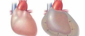
For a more clear idea of the pathological process, we can consider the main points of the pathophysiology of myocardial remodeling using the example of its structural changes after a heart attack. First of all, the shape of the left ventricle changed. If earlier its shape was an ellipse, now it looks more like a sphere. The thinning and stretching of the myocardium is very clearly observed, the area of necrosis of the area of the heart muscle often increases (even in cases where there were no repeated ischemic necrosis). Many other pathological disorders also appear that can lead to unpleasant consequences.
The interrelation of the processes during which a structural change develops in the heart muscle is obvious: first, the pressure increased, the heart tries to adapt in response to it, as a result, in direct proportion, the thickening of the ventricular wall occurs, and at the same time the weight of the muscle and some others increases, changes corresponding to a given state.
This example explains how myocardial remodeling occurs and why it can be dangerous and worsen the situation, increasing the risk of complications after a heart attack. That is why, after an attack, the patient undergoes a long period of rehabilitation; he is prescribed special medications (some of which are used continuously) to prevent the development of a relapse.
3Types of hypertrophy
Left ventricular hypertrophy
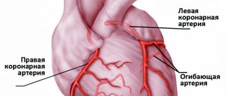
Concentric. Concentric hypertrophy of the left ventricular myocardium (concentric hypertrophy of the left ventricular myocardium) is characterized by uniform thickening of its walls. Such uniform thickening of the wall can lead to a decrease in the lumen of the chamber. Hence the second name for this type of hypertrophy is symmetrical. Most often, concentric LV hypertrophy develops due to pressure overload. Some pathological conditions and diseases, such as aortic stenosis and arterial hypertension (AH), lead to an increase in vascular resistance in the aorta. The left ventricle has to work harder to push all the blood into the aorta. This is where concentric LV hypertrophy develops.
Eccentric. Unlike the previous type, eccentric hypertrophy of the left ventricle is formed if the LV is overloaded with volume. Insufficiency of the mitral or aortic valve, as well as some other reasons, may lead to the fact that blood from the left ventricle is not completely expelled into the aorta. There remains some amount of it. The walls of the left ventricle begin to stretch, and its shape resembles an inflated balloon. The second name for this type of remodeling is asymmetric. With eccentric hypertrophy of the left ventricle, the thickness of its wall may not change, but the lumen, on the contrary, expands. Under such conditions, the pumping function of the left ventricle decreases.
The mixed type of hypertrophy most often occurs when playing sports. Individuals involved in rowing, speed skating or cycling may have this type of LV myocardial hypertrophy.
Separately, the authors highlight concentric remodeling of the LV myocardium. Its difference from concentric LVH is the unchanged mass of the LV myocardium and the normal thickness of its wall. With this type, there is a decrease in end-diastolic size (EDD) and LV volume.
How is it diagnosed and can the pathology be stopped?
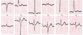
Diagnosis of this disease is carried out by taking an electrocardiogram of the heart. On it, if the geometry of the left ventricle of the myocardium changes, an increase in ST and a decrease in the R wave will be observed.
The development of myocardial remodeling can be prevented if hypertension is diagnosed in a timely manner (it is characterized by frequent upward pressure surges, headaches, and deterioration in general health).
Modern medicine proves that even existing pathology can be reduced with the help of medications and more. It is possible to reduce the thickness of the walls and reduce the mass of the left ventricle with the help of antihypertensive drugs.
Beta-blockers inhibit remodeling and improve the geometry of the left ventricle of the myocardium. In addition, on the first day after a heart attack, angiotensin-converting enzyme inhibitors are prescribed to prevent heart failure and prevent relapse. Nitrates, as well as calcium antagonists (they require a long course of therapy), are effective in limiting early post-infarction remodeling.
It is also important to reduce your intake of salt and pickles, follow a specially designed diet and take control of your own weight (prevent the formation of excess kilograms).
Concentric shape
Concentric myocardial remodeling
Concentric remodeling of the left ventricular myocardium is a fairly common finding that applies to patients with hypertension. The process begins with LV hypertrophy, which is manifested by an increase in the thickness of its wall. Changes in the septum are also noted. The interior space has not been changed.
It should be noted that the cause of LVH can be not only a persistent increase in blood pressure, but also other factors, such as:
- intense physical activity to which a person constantly subjects his body;
- sedentary lifestyle, often found among office workers;
- smoking, regardless of the number of cigarettes smoked;
- systematic alcohol abuse.
Thus, we can conclude that in order to prevent the start of the process of myocardial remodeling, it is necessary to diagnose hypertension or LVH as early as possible and begin their effective treatment. To do this, you need to study the symptoms that may indicate the presence of such diseases, these are:
- systematic increase in blood pressure;
- frequent headaches and dizziness;
- periodic tremors in the limbs;
- heart rhythm disturbances;
- difficulty breathing, shortness of breath;
- decreased performance;
- pain in the heart area.
If such signs occur, it is necessary to seek medical help and undergo a full examination, which allows you to obtain complete information about the state of your own health.
What is cardiac remodeling?
Remodeling is a phenomenon whose essence is to change the structure of an object. Changes in the structure and shape of the heart, including an increase in the weight of the left ventricular muscle and an increase in the size of the parts of the organ, which lead to a decrease in its functionality, are called myocardial remodeling. This process can occur rapidly, but more often it is long-term. With timely diagnosis, proper treatment, and elimination of the provoking factor, this process can be stopped and reversible.
2Formation of “perestroika”
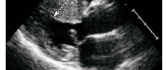
Scheme of the formation of two types of myocardial dysfunction
You can find the definition of hypertrophy as remodeling. These terms are synonymous with each other, although it is correct to say that hypertrophy is a particular remodeling. The second concept is broader. Remodeling means the process of changing an existing structure, rebuilding it, or adding something to it. Myocardial remodeling is a change in its geometric structure under the influence of a specific factor. Moreover, not only the structure is being rebuilt, but functional restructuring is also taking place.
The purpose of remodeling is to adapt the left ventricle to the established hemodynamic conditions, which often become pathological. With the constant influence of increased pressure on the LV myocardium, there is a response increase in the number of sarcomeres and the thickness of the heart cell (cardiomyocytes). As a result, the LV wall thickens, which occurs with concentric remodeling of the left ventricular myocardium. In the case of eccentric remodeling, the ventricle experiences volume overload. In this case, the cardiomyocytes are stretched, and the wall of the heart chamber decreases.
Remodeling
The following components are involved in the development of left ventricular (LV) myocardial remodeling:
- Myocardial cells are cardiomyocytes. Cardiomyoitis are highly differentiated structures. This means that these cells have lost the ability to divide. Therefore, in response to increasing physical activity (PE), the concentration of biologically active substances in the body increases: norepinephrine, angiotensin, endothelin, etc. In response to this, the number of sarcoplasmic contractile units in cardiomyocytes increases. Energy exchange processes begin to occur more intensively in the cell.
- Fibroblasts are components of connective tissue. While the myocardium thickens and hypertrophies, the vessels do not have time to provide such muscle mass with oxygen and nutrients. Oxygen requirements increase, but the vascular network remains at the same level. The LV myocardium enters a state of ischemia—oxygen starvation. In response to this, connective tissue components—fibroblasts—are activated. “Growing” with connective tissue, the myocardium loses its elasticity and becomes rigid. This circumstance entails a decrease in diastolic function of the left ventricle. In simple terms, diastolic dysfunction of the left ventricle (LV) appears.
- Collagen. In various diseases, in particular myocardial infarction, collagen, which ensures the relationship between cardiomyocytes, begins to weaken and disintegrate. The process of collagen formation does not keep pace with its breakdown in the first weeks of a heart attack. Then these processes are leveled out, and in place of weakened cardiomyocytes that have undergone necrosis during a heart attack, a connective tissue scar is formed.