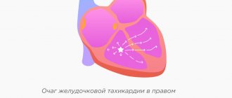
Ventricular tachycardia (VT) is a type of paroxysmal tachycardia (link to page). It is characterized by disturbance of sinus rhythm and an increase in the number of ventricular contractions. The cause of the disease is changes in the heart muscle or the conduction system of the ventricles of the heart, as a result of which the re-entry mechanism (circular arrhythmia) is realized, or some of the heart cells begin to exhibit increased electrical activity, which leads to accelerated work of the ventricles of the heart.
During an attack, the heart rate (HR) ranges from 140 to 220 beats per minute. Among all arrhythmias, VT is the most dangerous, because it can transform into a life-threatening arrhythmia - ventricular fibrillation. In addition, during an attack the ventricles contract frequently, but not fully. This can lead to insufficient blood supply to the brain and cause loss of consciousness.
Causes of ventricular tachycardia
In 98%, the cause of VT is a heart disease, in the remaining 2% the cause remains undetected. This pathology is called idiopathic.
Diseases that can lead to VT:
- myocardial infarction;
- post-infarction aneurysm;
- reperfusion arrhythmias;
- myocarditis;
- cardiosclerosis;
- cardiomyopathy;
- sarcoidosis;
- heart defects;
- genetic diseases
Let us note the provoking external factors influencing the development of VT:
- stressful situations;
- frequent psycho-emotional stress;
- increased physical activity;
- surgical intervention;
- drug overdose;
- hormonal imbalance in the body.
Main clinical symptoms
Paroxysmal ventricular tachycardia (VT)
More often against the background of gross chronic changes in the myocardium (post-infarction cardiosclerosis, rheumatism, etc.) or as a complication of acute myocardial infarction. May occur against the background of intoxication with cardiac glycosides, long QT syndrome (bidirectional fusiform ventricular tachycardia).
- As a rule – arterial hypotension;
- On auscultation of the heart, there is a cannon tone;
- ECG: rhythmic, extended (> 0.12 sec.) and deformed ventricular complexes with a frequency of 120-180 per minute, coupled or combined contractions, atrioventricular dissociation;
For differential diagnosis with supraventricular tachycardias with aberrant QRS complexes - drug tests (ATP or Lidocaine).
Symptoms of ventricular tachycardia
Symptoms depend on the duration of the attack and the complexity of the disorders. Sometimes VT is asymptomatic.
The main ailments include:
- cardiopalmus;
- pain and burning in the chest;
- severe dizziness;
- loss of consciousness;
- convulsions;
- weakness;
- anxiety or feeling of fear.
To be sure that your heart is working properly and to eliminate the cause of VT symptoms in a timely manner, consult our specialists. The Cardiology Center of the Federal Scientific and Clinical Center of the Federal Medical and Biological Agency has developed several heart examination programs. Cardiologists will select the right program for you and prescribe appropriate diagnostics.
Tachycardia with wide QRS complex
In case of paroxysm with a wide QRS complex (more than 0.12 s) and the impossibility of recording a transesophageal ECG, the following cannot be excluded:
• paroxysm of supraventricular tachycardia with blockade of the legs of the atrioventricular bundle (permanent or transient, frequency-dependent);
• paroxysm of supraventricular tachycardia, atrial fibrillation and flutter with impulse conduction along additional pathways.
In doubtful cases, wide complex tachycardia should be regarded as ventricular.
Diagnostics
If you feel a sudden increase in heart rate, you should immediately consult a doctor. After a physical examination, the patient is sent for an ECG to accurately determine the diagnosis.
As additional diagnostics use:
- daily ECG monitoring;
- EPI (electrophysiological study of the heart);
- ECHO;
- Coronary angiography is a radiopaque research method.
The doctor may order laboratory tests, the results of which will indicate concomitant pathologies.
Timely diagnosis is a fundamental factor in successful treatment of the disease. Do not ignore your ailments - seek help from the specialists of the Federal Scientific and Clinical Center of the Federal Medical and Biological Agency!
Risk of atrial fibrillation and flutter
Of particular danger is the occurrence of atrial fibrillation and flutter in patients with DPP and conduction of impulses along abnormal pathways anterogradely from the atria to the ventricles.
The risk of ventricular fibrillation can be increased, among other things, by the use of drugs that accelerate conduction through the AP. First of all, this applies to veralamil and digoxin, which precludes their use in the antidromic version of paroxysmal tachycardias. At the same time, the drugs of choice in the treatment of patients with paroxysmal tachycardia and widened QRS complex in WPW syndrome are drugs of class I (procainamide, disopyramide, propafenone) and class III (amiodarone).
It is often necessary to differentiate tachycardia with a wide QRS complex in WPW syndrome and paroxysm of ventricular tachycardia, which can be extremely difficult to do using an external ECG and requires transesophageal ECG diagnostics.
A very high ventricular rate (more than 200 per minute), the occurrence of tachycardia paroxysms in childhood and adolescence (often in patients without structural changes in the myocardium), and the presence of tachycardia attacks in close relatives may indicate to the doctor the possibility of the existence of DPP.
Prevention of ventricular tachycardia
When symptoms are detected, immediate diagnosis is important in order to eliminate the cause of the disease and treat it. In most cases, prevention is secondary and consists of preventing relapse. Therefore, an important part of prevention is regular examination of the cardiovascular system. Specialists of the Cardiology Center of the Federal Scientific and Clinical Center of the Federal Medical and Biological Agency conduct comprehensive heart checks; you can find out about the programs here.
To prevent the occurrence or recurrence of ventricular tachycardia, it is important to follow the doctor's recommendations and follow all his instructions.
Additional preventive measures include:
- regular blood pressure monitoring;
- body weight control;
- proper nutrition: exclude fatty, fried, spicy, salt;
- complete cessation of bad habits;
- exercise therapy and moderate physical activity.
emergency care for paroxysmal tachycardia
Emergency care for paroxysmal tachycardia
Supraventricular tachycardia.
QRS duration is normal or less than 0.08 seconds. Techniques of vagal irritation or vagotonic reflex tests are used only for supraventricular paroxysmal tachycardia with normal QRS complexes. They are not used in young children and are most effective in the first 20 minutes after an attack. It is preferable to prescribe to children over 3 years of age.
If reflex methods are ineffective (if the child’s condition does not require extreme measures), for children over 3 years of age: crushed and in a sufficient amount of liquid, seduxen 1\3-1 tablet, isoptin 1\3-1 tablet. (1 mg/kg) and 1-2 tablets. panagin, after an hour can be repeated in the same doses. The child can be put to bed, creating a calm environment (protective mode).
It is possible to combine verapamil (1-2 mg/kg) and the beta-blocker pindolol (0.25 mg/kg) in the form of crushed tablets. The effect is observed on average after 10 minutes in 75% of patients, and the drugs are well tolerated.
Propranolol can be administered orally in a single dose of 1 mg/kg in 3 doses with an interval of 2 hours. But it is more advisable to use propranolol (1-2 mg/kg) and diltiazem (voltage-dependent calcium channel blocker) (1-2 mg/kg), which first reduce the severity of tachycardia, and then, on average, stop the attack after 30-40 minutes.
Pindolol - indications are the same as propranalol, if administered 0.1-0.2 mg/kg/day slowly.
If there is no positive dynamics or the child’s clinical condition worsens, as well as in young children, it is necessary to start with intravenous administration.
1. Isoptin (verapamil) 2.5% - 0.1-0.2 mg/kg IV in 20 ml of 10% glucose solution in combination with seduxen and 2-5 ml of panangin. Since the effect is short-term (40-60 minutes), if there is no effect, repeat after 30 minutes. Can be combined with sibazon and 2-5 ml of panagin.
2. Adenosine monophosphate (AMP) or phosphadene 2% solution 0.1 mg/kg for a very fast bolus (2-3 sec), diluted with saline to avoid sequestration of adenosine in red blood cells. The dose can be doubled and repeated again until the attack stops or side effects appear (redness, shortness of breath, chest pain, bradycardia, irritability, but usually disappear within 1-2 minutes). The maximum single dose should not exceed 12 mg or 0.3 mg/kg. Can be used for all types of tachyarrhythmias. It has a direct inhibitory effect on the AV node.
3. ATP (phosphobion ) up to 6 months 0.5 ml, 6-1 years -0.7 ml, 1-3 years -0.8, 4-7 years -1.0, 8-10 years 1.5 ml, 11-14-2.0 ml. administer without dilution quickly (2-3 seconds). Similar action with adenosine.
4. Procainomide (novocainomide) – used in the absence of heart failure, arterial hypotension, blockades, and simultaneous use of digoxin. 10% -5.0 -0.15-0.2 ml/kg IV together with 1% mesaton solution - 0.1 ml/year of life (to prevent a sharp drop in blood pressure, especially against the background of heart failure. It also inhibits antegrade conduction in the AV node ).
5.0.1% solution of anaprilin (obzidan), propranolol - 0.01-0.02 mg/kg, very slowly in a stream.
6. Ajmaline (gilurythmal) iv slowly 2.5% single dose 1 mg/kg in 20 ml of saline solution.
7. If other drugs are ineffective, a 5% solution of cardarone (amiodarone) 5 mg/kg or 0.1 ml/kg in 150 ml of a 5% glucose solution blocks K channels, to a lesser extent Ca channels, inhibits a and b receptors.
Ventricular tachycardia.
Ventricular QRS is wide for ages greater than 0.08 sec.
1. Lidocaine 1% - 1 mg/kg IV for 2 minutes, if there is no effect, can be repeated after 5-10 minutes. Then 2 mg/min intravenously in the first 12 hours, then 1 mg/min for 12 hours. The second drug is procainomide, which is administered intravenously as a bolus of 100 mg every 5 minutes until the gastrointestinal tract is eliminated or a total dose of 10-20 mg/kg is achieved. Monitoring blood pressure and ECG. Then administer 2 mg/min intravenously over several hours.
2. If there is no effect: novocainomide (procainomide) 10% - 5.0 -0.15-0.2 ml / kg IV in a slow stream along with a 1% solution of mezaton - 0.1 ml / year of life (to prevent a sharp drop in blood pressure, especially against the background of heart failure. It also inhibits antegrade conduction in the AV node).
3. If the first two are ineffective - Ornid (bretylium tosylate) - sympatholytic -5% -1.0 - 5-10 mg/kg. in 50 ml of 5% glucose for 20 minutes.
4. If malignant gastric tract is resistant to other drugs, a 5% solution of cardarone (amiodarone) 5 mg/kg or 0.1 ml/kg in 150 ml of 5% glucose solution is indicated - it blocks K channels, to a lesser extent Ca channels, inhibits a and b receptors.
5.0.1% solution of anaprilin (obzidan), propranolol - 0.01-0.02 mg/kg, very slowly in a stream. If the attack does not stop for a long time (more than 24 hours), signs of circulatory failure increase, the neck veins swell, pallor and cyanosis increase, then defibrillation is indicated. Carrying out cardioversion (a method of treating tachyarrhythmias using a defibrillator). When paroxysmal tachycardia is combined with heart failure, IV digoxin 0.025% -0.03-0.05 mg/kg/day for 3 injections - rapid saturation. Half the daily dose in the first administration. Digoxin is combined with the administration of propronalol and verapamil, but propronalol and verapamil should not be given together due to a sharp drop in blood pressure and the risk of cardiac arrest.
Diagnostic measures
- Taking anamnesis (simultaneously with diagnostic and therapeutic measures);
- Examination by an emergency medical technician (paramedic) or a medical specialist from a visiting emergency medical team of the appropriate profile;
- Registration of an electrocardiogram, decoding, description and interpretation of electrocardiographic data;
- Monitoring of electrocardiographic data;
- Pulse oximetry;
- General thermometry.
For unstable hemodynamics:
- Diuresis control;
For anesthesiologists and resuscitators:
- Control of central venous pressure (in the presence of central venous access).
Therapeutic measures
Paroxysmal ventricular tachycardia with stable hemodynamics and absence of clinical manifestations of pulmonary edema:
- Providing a medical and protective regime;
- Horizontal position;
- Inhalation administration of 100% O2 on a constant flow through a mask or nasal catheters;
- Catheterization of the cubital or other peripheral veins or installation of intraosseous access;
- Option I:
- Amiodarone (Cordarone) – 5 mg/kg IV (intraosseous) bolus slowly over 3 minutes (maximum 300 mg);
When stopping VT:
- Amiodarone (Cordarone) – 150 mg, intraosseous drip or infusion pump, at a rate of 1 mg/min., on site and during medical evacuation;
Option II:
- Lidocaine – 1.5 mg/kg IV (intraosseous) bolus slowly (up to 120 mg) +
- Lidocaine – 1.5 mg/kg, intraosseous drip or infusion pump, at a rate of 2 mg/min., on site and during medical evacuation;
If there is no effect:
For cardiologists and resuscitation doctors:
- EIT: initial discharge energy – 200 J (monophasic discharge), 120-150 J (biphasic discharge), with preliminary analgesia and sedation (Narcotic analgesic IV (intraosseous) or Ketamine IV (intraosseous) + Diazepam IV ( intraosseous);
- With polymorphic ventricular tachycardia (pirouette type):
- Magnesium sulfate – 2500 mg IV (intraosseous) slow bolus;
If there is no effect after 10 minutes:
- Magnesium sulfate – 2500 mg IV (intraosseous) slow bolus;
If there is no effect:
For cardiologists and resuscitation doctors:
- EIT: initial discharge energy – 200 J (monophasic discharge), 120-150 J (biphasic discharge), with preliminary analgesia and sedation (Narcotic analgesic IV (intraosseous) or Ketamine IV (intraosseous) + Diazepam IV ( intraosseous);
- Additional scope of treatment measures for the underlying disease;
- Medical evacuation (see “Scope of tactical measures”).
Paroxysmal ventricular tachycardia with unstable hemodynamics or in the presence of clinical manifestations of pulmonary edema:
- Providing a medical and protective regime;
- Horizontal or anti-shock position;
- Inhalation administration of 100% O2 at a constant flow through a mask or nasal catheters, in the presence of moist rales in the lungs: with alcohol vapor;
- Catheterization of the cubital or other peripheral veins or installation of intraosseous access;
- In the absence of clinical manifestations of pulmonary edema:
- Sodium chloride 0.9% – IV (intraosseous), drip, at a rate of 10 ml/kg/hour, under auscultatory control of the lungs, on site and during medical evacuation;
- For cardiologists and resuscitation doctors:
- EIT: initial discharge energy - 200 J (monophasic discharge), 120-150 J (biphasic discharge), with preliminary analgesia and sedation (Narcotic analgesic IV (intraosseous) or, in the absence of clinical manifestations of pulmonary edema, Ketamine IV (intraosseous ) + Diazepam IV (intraosseous);
If there is no effect:
- EIT – 360 J (monophasic discharge), 200 J (biphasic discharge);
If there is no effect:
- EIT – 360 J (monophasic discharge), 200 J (biphasic discharge);
If there is no effect from triple EIT:
- Drug defibrillation + EIT,
Option I:
- Amiodarone (Cordarone) – 300 mg IV (intraosseous) (intraosseous) slow bolus + EIT – 360 J (monophasic shock), 200 J (biphasic shock) + every 2 minutes: EIT – 360 J (monophasic shock), 200 J (biphasic discharge);
h/w 15 minutes again:
- Amiodarone (Cordarone) – 150 mg IV (intraosseous) bolus slowly + EIT – 360 J (monophasic discharge), 200 J (biphasic discharge), + every 2 minutes – EIT – 360 J (monophasic discharge), 200 J (biphasic discharge);
When stopping VT:
- Amiodarone (Cordarone) – 150 mg, intraosseous drip or infusion pump, at a rate of 1 mg/min., on site and during medical evacuation;
Option II:
- Lidocaine – 1.5 mg/kg IV (intraosseous) slow bolus + EIT – 360 J (monophasic discharge), 200 J (biphasic discharge) +
- Lidocaine -1.5 mg/kg, intraosseous drip or infusion pump, at a rate of 2 mg/min. + EIT – 360 J (monophasic discharge), 200 J (biphasic discharge) – every 2 minutes until effect,
When stopping VT:
- Lidocaine -1.5 mg/kg, intraosseous drip or infusion pump, at a rate of 2 mg/min., on site and during medical evacuation;
If it is impossible to carry out EIT:
For teams of all profiles:
- Dopamine – 200 mg IV (intraosseous) by drip or infusion pump, at a rate of 5 to 20 mcg/kg/min., on site and during medical evacuation or, and
- Norepinephrine – 4 mg IV (intraosseous) by drip or infusion pump, at a rate of 2 mcg/min., on site and during medical evacuation or, and
- Adrenaline – 1 – 3 mg IV (intraosseous) by drip or infusion pump, at a rate of 2 to 10 mcg/min., on site and during medical evacuation;
If blood pressure increases >100 mm Hg: –
- Medical defibrillation without EIT;
- If there are clinical manifestations of pulmonary edema:
- Additional scope of treatment measures according to the protocol “Left ventricular failure”;
- Additional scope of treatment measures for the underlying disease;
- Medical evacuation (see “Scope of tactical measures”).
