We recommend reading the article on coronary circulation. From it you will learn about the diagram of the coronary circle, physiology, regulation of the small coronary circle, and research methods. And here is more information about hypoxemia and hypoxia.
What is it, its color, main characteristics and composition
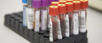
Venous blood is that which flows from the organs to the heart, and from it enters the lungs. It takes in carbon dioxide and metabolic products. It is used for testing and medications are also injected into it through intravenous injections. There are differences in the main characteristics and composition between the blood in the arterial and venous beds; they are presented in the table.
It is important to note that the maximum difference between arterial and venous blood concerns gas composition, pressure and flow rate. All other physical and chemical properties differ only slightly.
Functions
Main functions of venous blood:
- removal of metabolic products (cleansing);
- transfer of absorbed nutrients from the intestine to the liver through the portal vein (transport);
- removal of excess hormones, salts (maintaining balance, equilibrium, homeostatic);
- delivery of carbon dioxide to the lungs for removal from the body (respiratory).
What is venous blood rich in?
Venous blood is rich in carbon dioxide; it also contains metabolic end products, toxins and microbial remains. Because of this, it is possible to identify the main diseases:
- infections;
- metabolic disorders (for example, atherosclerosis, diabetes mellitus);
- immune, allergic, autoimmune pathologies;
- blood diseases due to changes in the level of red blood cells, leukocytes, platelets;
- dehydration;
- risk of increased thrombus formation and blockage of veins and arteries;
- liver and kidney dysfunctions;
- hormonal disbalance.
Elements of the human circulatory system containing venous blood
Venous blood is contained in the following elements of the circulatory system:
- subcutaneous and deep venous vessels;
- venous networks of internal organs, brain;
- large vena cava (superior and inferior), carrying blood to the right atrium;
- the pulmonary artery emerging from the right ventricle and its branches in the lungs.
Artery through which venous blood flows
Venous blood flows through the pulmonary artery. This name is due to the fact that all the vessels leaving the heart are called arteries, and those coming to it are called veins. Therefore, in the veins of the lungs there is an arterial vein, rich in oxygen, and in the arterial network - carbon dioxide.
Which parts of the heart contain venous blood?
Venous blood is found in the right atrium and right ventricle of the heart. If there is a defect in the septum between the right and left parts, then a mixture of arterial and venous occurs.
Why are tests taken from a vein?
All metabolic products, hormones and toxins accumulate in the veins. Therefore, by venous blood it is possible to determine diseases of internal organs and the brain, which are manifested by changes in the composition of cells or plasma (liquid part). A puncture of a vein causes slight bleeding, which can be stopped very quickly by squeezing the vessel. If there is even slight damage to the artery, a tourniquet will be needed.
Arterial and venous blood
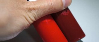
Differences between arterial and venous blood: dark blood flows from the veins, it is thicker, it clots faster, the bleeding is less intense, the stream is smooth and not strained. Venous blood is more suitable for laboratory tests than capillary blood from a finger. It flows through the pulmonary arteries, and in the capillaries of the alveoli it turns into arterial artery.
Which one is darker
Venous blood is darker. Its color depends on the form of hemoglobin. In the arterial blood, it is combined with oxygen (oxyhemoglobin), which gives it a bright scarlet color. The venous contains both oxyhemoglobin and 2 other forms:
- reduced (gave oxygen to cells, but has not yet added carbon dioxide);
- carboxyhemoglobin (a compound with carbon dioxide).
Most of the latter pigment, so the color becomes dark cherry.
Capillary and venous blood: differences
The main differences between capillary blood (from a finger) and venous blood are the contents:
- cells, especially platelets (higher in the venous), leukocytes (higher in the capillary);
- glucose (higher in the veins).
Analysis from a vein is considered more accurate, since it does not contain impurities of tissue fluid or surface epithelium of the skin. Also, circulatory disorders, vascular spasm, and fever can affect the indicators of capillary blood tests. For many types of laboratory diagnostics, a sufficient volume of material is needed, and up to 0.5 ml can be taken from a finger.
Therefore, capillary blood can be used only at the first stage of the examination. It will give reliable results only when determining hemoglobin, red blood cells and ESR. For biochemical and immunological analysis, studies of hormonal levels, as well as, if necessary, an in-depth study of cellular composition, blood from a vein is needed.
Venous system of the lower extremities
The main task of the venous system is to ensure complete outflow of venous blood from the lower extremities at rest and during physical activity.
This process is carried out through three types of veins: superficial, deep and communicating (perforating, commissural, perforating).
Superficial veins.
Superficial veins lie directly under the skin and provide the outflow of venous blood (which is approximately 30% of the total volume of venous blood) from the superficial layers of the skin and subcutaneous fat.
Deep veins - located in the thickness of the muscles, accompany in pairs the arteries of the same name and their large branches. They receive more than 70% of the total volume of venous outflow.
The role of an additional “pump” that lifts this amount of blood to the level of the heart in the body is performed by a powerful group of muscles of the thigh and lower leg. While walking, the muscles contract and act on the deep veins passing through them, and the blood is pushed upward. This co-operative anatomical formation is called the muscular-venous pump.
Communicating veins connect the superficial and deep veins with each other, and also carry out the redistribution of venous blood between them.
To maintain the direction of blood flow, the veins are equipped with valves. Normally, their work ensures unidirectional blood flow to the heart. The valves are folds (similar to pockets), which are located in pairs on the inner surface of the vein wall every 8-9 cm. When reverse blood flow occurs, the pockets are filled, as a result, the valve flaps close and the lumen of the vessel is blocked.
The valve system of the communicating veins is designed in such a way that when the pressure in the superficial veins increases, excess blood is drained into the deep vein system. Due to this, the “weaker” and more prone to expansion superficial veins are protected from overflow even with a significant increase in venous pressure.
The largest number of communicating veins is located in the lower third of the leg. As can be seen in Figure 4.5, muscle tendons are mainly located here, and muscle mass is weakly expressed. To maintain the unidirectional flow of venous blood, the largest number of valves are located here in the veins (more than half of the total number of all valves of the lower limb).
Normally, the presence of a pressure difference between adjacent sections of the vein, the coordinated work of the muscular-venous pump and the valve apparatus of the veins play an important role in the return of venous blood and the unidirectionality of blood flow.
During active life (a person's waking time is approximately 2/3 of the day), most of the circulating blood (60 - 70%) is below the level of the heart, which creates certain difficulties in its return to the heart. Therefore, the greatest problems arise in a person’s leg veins. Exposure to various factors can provoke the development of abnormalities, which later transform into diseases of the veins and lymphatic vessels.
- Back
- Forward
- Are you here:
- home
- Express orthotics
- 4.1 Human cardiovascular system
InterpretationTranslation
Venous blood Venous donor blood.
Deoxygenated blood
- blood returning to the heart through the veins. With the exception of the blood in the pulmonary veins, venous blood is deprived of oxygen and enriched with carbon dioxide as a result of tissue gas exchange. Venous blood is usually warmer than arterial blood, has a lower pH, contains less glucose and other nutrients, and more metabolic end products (urea, etc.).
Venous blood is obtained by venipuncture. Most medical laboratory blood tests are performed with venous blood.
Venous blood has a dark red color with a bluish tint.
Category:
- Blood
Help students solve problems
Incomplete separation of arterial and venous blood
Incomplete separation of arterial and venous blood occurs with heart defects and large vessels , as well as lung diseases:
- atrial septal defect;
- a hole in the septum between the ventricles;
- collapse of the walls (closing) of the alveoli of the lungs (atelectasis), filling with fluid (edema, pneumonia);
- fistula between a vein and an artery (congenital, less commonly acquired);
- transposition of the great vessels (aorta and pulmonary artery change places);
- underdevelopment of the heart chambers (two- and three-chamber);
- open arterial (ductus Botallus, connects the aorta and pulmonary artery).
Dark blood from a vein: what does it mean?
The dark color of blood from a vein means that the organ from which it flows is actively functioning. The higher the rate of metabolic processes, the more oxygen is absorbed from the blood, which means its color will be darker.
We recommend reading the article about venous congestion in the legs. From it you will learn about the causes and symptoms of the pathology, conservative and surgical treatment.
Stagnation
The presence of venous blood in vessels and organs for a long time is called stagnation. This condition can have varying degrees. Venous blood can accumulate near the walls of blood vessels and in organs in small quantities or almost completely displace a separate section of the circulatory system.
When stagnation occurs, nutrients and oxygen cease to flow into individual vessels and organs. Gradually, the skin may become blue and painful when you press on certain areas of the skin. Stagnation of venous blood provokes inflammatory and purulent processes in certain areas of the body and can cause inflammatory processes in organs.
Symptoms and signs
Stagnation of venous blood does not immediately manifest symptoms.
The following symptoms gradually develop:
- headache, especially in the morning;
- pain when pressing and moving in certain parts of the body;
- the appearance of bluish integuments;
- the appearance of slight congestion in the ears;
- development of nervous diseases.
Causes
Venous blood has the property of carrying viruses, bacteria, and cellular metabolic products for their removal from the body and always flows from the organ.
Due to this feature, stagnation of venous blood can lead to the development of serious disorders in the body, the causes of which may be:
- Injury due to blows and bruises. Hemorrhages and stagnation of venous blood appear not only after injury, but also as a result of improper fusion of vessels in which venous blood can stagnate.
- Blood clots or complete or partial blocking of vessel walls with blood clots disrupts the normal flow of venous blood, the pressure of which is not high. As a result, venous stagnation of blood occurs in the area with the thrombus.
- A sedentary lifestyle , in which the pressure in the pelvis increases as a result of prolonged sitting. Increased pressure in the lower abdomen in the presence of thick blood causes venous stagnation and serious health problems (hemorrhoids, varicose veins).
- Pinching certain areas of the body when wearing tight shoes, tight clothing) noticeably narrows the blood vessels in the compressed parts of the body. Circulation in these areas is disrupted and stagnation occurs.
- Neoplasms of any nature (malignant and benign) cause circulatory problems when they grow large. As a result of tissue compression, the blood circulation process is disrupted and venous stagnation occurs in certain areas of the vessels.
- Varicose veins change the course of the vessel, creating irregularities and depressions. In such places near the walls, there may be a slight stagnation of venous blood, which also provokes pain and discomfort.
- Congestion of the brain develops due to disorders of the blood vessels inside the brain. Damaged tissues swell and become inflamed, and oxygen starvation and congestion form inside the head.
- Stasis of the lungs or lower extremities , resulting in swelling and a marked increase in the size of the lungs and vascular tissues of the lower extremities. Blood circulation is disrupted, and venous blood stagnates in the tissues.
- Hyperemia of the kidneys or their significant thickening and enlargement provokes spasm and low blood flow to the organ.
Treatment methods
Venous blood can stagnate in different parts of the body and human organs, so treatment of stagnation is carried out using various methods, which include:
- review of the nutrition system and the use of dietary products, cessation of tobacco and alcohol;
- physical exercises in the form of race walking, combined with a contrast shower, help restore blood circulation;
- surgical intervention, which involves removing the damaged area, growth and restoring the normal functioning of blood vessels;
- drug therapy includes:
- ointments and gels for topical use that help restore blood microcirculation and promote vascular restoration. These include heparin ointment, troxerutin gel, venen-gel. Ointments and gels are applied to the damaged area of the body 2-3 times a day with massaging movements for 5-7 days;
- taking medications that help restore the condition of blood vessels and blood microcirculation. Such drugs relieve symptoms of heart failure and help eliminate symptoms of varicose veins. Curantil relieves venous blood stagnation by restoring the blood supply to damaged tissues. The drug is taken 1 tablet. 3 times a day before meals for 30-60 days;
- Taking diuretics helps accelerate blood flow and remove all harmful substances from the body along with the fluid. Furasemide also helps remove sodium and lower blood pressure. It is taken for stagnation, 1 tablet. 2-3 times a day for 7 days.
- taking antithrombosis drugs (warfarin, Plavix), which help clean blood vessels and restore blood circulation. Plavix is taken 1 capsule. 2 times a day for 7-10 days.
- taking antispasmodics allows you to relieve spasm of cerebral vessels and restore blood flow and remove congestion in the vessels of the head (no-spa, drotaverine, cordaferon). No-shpu during stagnation, take 1-2 tablets. 2 times a day for 2 weeks.
Traditional methods
Venous blood passes through the vessels of the circulatory system, therefore, to treat stagnation, traditional methods are often used, which have an effect on the circulatory system and the condition of the walls of blood vessels.
Folk remedies are used only after consultation with a specialist and in combination with other treatment methods. The following are used as folk remedies:
Sweet clover decoction
A decoction of sweet clover leaves helps restore blood circulation. It promotes the restoration of blood vessels and helps cleanse the blood of excess cellular metabolic waste products.
Cooking steps:
- To prepare the decoction 2 tbsp. l. dry sweet clover leaves combined with 1 tbsp. hot water. Then the mixture is boiled in a water bath for 20 minutes and filtered through a sieve.
- It is recommended to take 100 ml of the finished decoction. morning and evening on an empty stomach.
Nettle juice
Nettle juice helps restore the condition of blood vessels, improves blood circulation and has anti-inflammatory properties.
Cooking steps:
- Nettle leaves and stems are passed through a juicer or crushed in a blender and filtered.
- Store fresh nettle juice in the refrigerator and drink 100 ml. 3-4 times a day.
Grape juice
Fresh grape juice has the ability to strengthen the walls of blood vessels. It cleanses the walls of blood vessels and has diuretic properties.
Drink 1 tbsp of freshly squeezed grape juice. 3 times a day for 20-30 days.
Useful video
Watch the video about the circulatory system:
Pediatrician consultation / Svetlana, Tula December 3567, 2020
Hello, my daughter is 5 years old. Today I cut my finger with a new book until it bled. She came up to me and I saw that the blood was very light. Usually much brighter. What tests need to be done to rule out pathologies? I’m worried because she was recently sick, she had a sore throat and snot. I donated blood in August and it was normal. I’m also feeling fine, only recently I’ve had cold hands and feet, but the temperature hasn’t risen
On the Ask a Doctor service, you can consult a pediatrician on any problem that concerns you. Expert doctors provide consultations around the clock and free of charge. Ask your question and get an answer immediately!
Answers from doctors Complain Valeria Bychenko, December 29, 2020 Obstetrician, Gynecologist, Pediatric gynecologist Hello. Get a routine CBC and ferritin test. Light blood is a sign of anemia Complain Svetlana, December 29, 2021 Client Valeria, thank you. Can this be done after the holidays? She will just need to have a mantu done Complain Valeria Bychenko, December 29, 2021 Obstetrician, Gynecologist, Pediatric gynecologist You can. Now add beef, pomegranate (you can juice it), and green apples to your food. You can also give vitamin C (yellow tablets) Complain Albina Alimova, December 29, 2020 Pediatrician Complain Svetlana, December 29, 2021 Client Albina, curled up quickly. We will now take a blood test only after the holidays Complain Albina Alimova, December 29, 2021 Pediatrician Attach the results here. We will look Complain Svetlana, December 29, 2020 Client Albina, thank you. But now it’s only 12 hours Complain Albina Alimova, December 29, 2021 Pediatrician Good) The most important thing is don’t worry or worry. Maybe the cut was minor and there was little blood, which is why it seemed so to you. After ng, calmly take the tests) Complain Adele Syzdykova, December 29, 2021 Obstetrician, Gynecologist, Pediatric gynecologist Complain Svetlana, December 29, 2020 Client Adele, thank you. It’s just that I also recently cut my finger, I had a bright and thick one Complain Adele Syzdykova, December 29, 2020 Obstetrician, Gynecologist, Pediatric gynecologist Of course, you can understand, my little daughter. In any case, don't worry. Get tested on the 12th and rule out all pathologies. The most important thing is that the baby feels good, her appetite and vigor are in order. Complain Svetlana, December 29, 2021 Client Adele, she has problems with her appetite, she sometimes eats, sometimes she doesn’t. I don't know what to feed. Asks for sweets, bread Complain Adele Syzdykova, December 29, 2021 Obstetrician, Gynecologist, Pediatric gynecologist Which, by the way, may well lead to anemia. I think she is already manipulating you here. Try not to force her to eat, don’t focus on the food, don’t touch it if she doesn’t eat, but don’t give her sweets with bread. Complain Ekaterina Smirnova, December 29, 2021 Pediatrician, Pulmonologist Hello. Isn't the child himself pale? Are your lips pink? Complain Svetlana, December 29, 2021 Client Ekaterina, yes, sort of. Only heels underwear. But she has been like this since birth Complain Ekaterina Smirnova, December 29, 2021 Pediatrician, Pulmonologist Then your child does not have any pathology. It is impossible to cut deeply with a book, so it damaged the upper layers of the skin. And damage to the upper layers leads to abundant release of transudate (light liquid), which is why the blood is light. Remember what happens with an abrasion, in which the surface of the skin is slightly damaged (light pink water is released (this is transudate). It’s the same story. If the cut were deeper, then you would see the real color of the blood. Don’t look for pathologies in a child, there are none. Yours sincerely. Complain Maya Stepanova, December 29, 2021 Pediatrician Good evening, Svetlana! It is necessary to take a General blood test, blood tests with markers of anemia. Perhaps the cut was very superficial, and it was the discharge of ichor. Only an examination will give an understanding situation. Be healthy! + 1 Complain Svetlana, December 29, 2021 Client Maya, yes, there was less than a drop of blood Complain Zoya Zelenina, December 29, 2020 Anesthesiologist-reanimatologist, Pediatrician Clinical blood test to exclude anemia Complain Daria Dautbaeva, December 29, 2020 Pediatrician Hello! Perhaps the paresis was not so deep or oblique that it affected very small vessels, hence the color saturation is different. But poor appetite and pickiness in eating are one of the signs of anemia, so an examination makes sense! Complain Aida Karabekyan, December 29, 2021 Pediatrician, Ultrasound Doctor Hello! Tomorrow you can take a cytological test for your blood test and ferritin in any private center and have time to post the result here. Complain Evgeny Khodyrev, December 30, 2021 Pediatrician Complain Anna Rudik, December 30, 2020 Gastroenterologist, Therapist, Hematologist Hello. You need to take a general blood test and ferritin. If there are deviations, you will need to start taking iron supplements. Similar questions on the topic The embryo is not visible 7 answers December 10, 2021 Alena, Moscow The question is closed What should I do if I haven’t found the answer to my question?
If you have a similar or similar question, but you have not found the answer to it, get your 03 online consultation from an expert doctor.
If you want to get a more detailed consultation with a doctor and solve the problem quickly and individually, ask a paid question in a private personal message. Be healthy!
What determines the color of blood?
The red color of blood can have different shades. Oxygen carriers, i.e. red blood cells (erythrocytes), are a shade of red depending on hemoglobin, an iron-containing protein found in them that can bind with oxygen and carbon dioxide to carry them to the desired location. The more oxygen molecules connected to hemoglobin, the brighter the red color the blood is. That’s why arterial blood, which has just been enriched with oxygen, is so bright red. After the release of oxygen to the cells of the body, the color of the blood changes to dark red (burgundy) - such blood is called venous.
Of course, the blood contains other cells besides red blood cells. These are also leukocytes (white blood cells) and platelets. But they are not in such significant quantities compared to red blood cells as to affect the color of the blood.
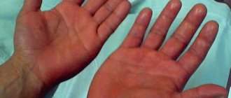
Blood color in anemia and cyanosis
With anemia (not enough hemoglobin or red blood cells), the blood can be said to be a paler red color, although this can only be seen by a specialist under a microscope. This is because when hemoglobin is not bound to oxygen, the red blood cells appear smaller and paler.
When the blood, due to health problems, does not carry enough oxygen and there is little oxygen in it, this is called cyanosis (cyanosis). That is, there is hemoglobin in the blood, but it is not associated with oxygen. A manifestation of cyanosis is the acquisition of a bluish tint by the skin and mucous membranes. The blood remains red, but even arterial blood has a color similar to the color of venous blood in a healthy person - with a blue tint. The skin, under which the vessels pass, which under normal conditions transport bright scarlet blood rich in oxygen, becomes blue in appearance.
But with anemia, the symptoms of cyanosis may not even be visible, because there is too little hemoglobin to affect the color of the skin and mucous membranes, and they are simply pale. In this case, external cyanosis will begin to appear only when the amount of reduced (without oxygen) hemoglobin becomes more than half of its total amount.
dark red, bright red, composition of human blood
Why is venous blood darker than arterial blood, and how to determine the type of bleeding
Arterial bleeding is one of the most dangerous bleedings, posing a direct threat to human life. This is primarily due to the fact that blood loss is high and intense. Therefore, it is important to know its main signs and first aid rules.
the color of the blood is bright scarlet, its consistency is liquid, it does not flow out of the wound, but flows out in a powerful stream, similar to a stream in a fountain. There is always a pulsation that occurs in time with the contraction of the heart muscle. Since blood comes out very quickly, a person may experience vasospasm and loss of consciousness.
First aid rules will vary depending on where the injury is located and which artery was damaged:
- First of all, it is necessary to apply a tourniquet that will prevent blood loss. Before fixing it, it is important to press the injured artery to the bone, above the place where the blood is leaking. If the shoulder is wounded, the fist is placed in the armpit, and the arm is pressed against the body; if the forearm is wounded, place any suitable-sized object in the elbow crease and bend the arm as strongly as possible in this joint. If the thigh is wounded, the artery is pinched with a fist in the groin area; if the lower leg is wounded, a corresponding object is placed in the popliteal area and the leg is bent at the joint.
- The limb should be raised and a cloth should be placed under the tourniquet. When there is no rubber band at hand, it can be replaced with an ordinary bandage or strip of fabric. For a tighter fixation, you can use a regular stick.
- It is important not to leave the tourniquet on the limb too long; it must be removed after 1 – 1.5 hours, depending on the time of year. It is best to record the time of its application on paper and place it under the bandage. This must be done to prevent tissue death and amputation of the limb.
- When the time for wearing the tourniquet has expired and the victim is not hospitalized, it is necessary to loosen it for a few minutes. In this case, the wound should be closed with your hands using a clean cloth.
- Deliver the victim to a medical facility as quickly as possible, where he will receive qualified assistance.
The rules for assistance in the event of arterial bleeding from the feet and hands differ. In this case, there is no need to apply a tourniquet. It is enough to bandage the injured area and raise it higher.
When arteries such as the subclavian, iliac, carotid or temporal are injured, the blood is stopped using tight wound tamponade. To do this, either sterile cotton wool or sterile napkins are placed in the damaged area, then a layer of bandage is placed on top and wrapped tightly.
Why is venous blood darker than arterial blood, and how to determine the type of bleeding
Do you know what color the blood in the veins is? The shade of biological fluid determines the presence of hemoglobin in red blood cells (erythrocytes). The blood circulating through the arteries, as already mentioned, is scarlet.
This is due to the high concentration of hemoglobin (in humans) and hemocyanin (in arthropods and mollusks), enriched with various nutrients.
Venous blood has a dark red tint. This is due to oxidized and reduced hemoglobin.
At the very least, it is unreasonable to believe the theory according to which the biological fluid circulating through the vessels is bluish in color, and when injured and in contact with air due to a chemical reaction, it immediately turns red. It is a myth.
Veins can only appear bluish, this is due to the simple laws of physics. When light hits the body, the skin reflects some of all the waves and therefore looks light or dark (this depends on the concentration of the coloring pigment).
You know what color venous blood is, now let's talk about the composition. You can distinguish arterial blood from venous blood using laboratory tests. Oxygen tension - 38-40 mmHg. (in venous), and in arterial blood - 90. The content of carbon dioxide in venous blood is 60 millimeters of mercury, and in arterial blood - about 30. The pH level in venous blood is 7.35, and in arterial blood - 7.4.
The outflow of blood, carrying away carbon dioxide and products formed during metabolism, is carried out through the veins. It is enriched with useful substances that are absorbed into the walls of the gastrointestinal tract and produced by vital substances.
Now you know what color the blood is in the veins, you are familiar with its composition and functions.
Blood flowing through the veins overcomes “difficulties” during movement, which include pressure and gravity. That is why, in case of damage, biological fluid flows in a slow stream. But if the arteries are injured, blood spurts out like a fountain.
The speed at which venous blood moves is much less than the speed at which arterial blood moves. The heart pumps blood out under high pressure. After it passes through the capillaries and becomes venous, a decrease in pressure to ten millimeters of mercury is noted.
Venous bleeding is characterized by the effusion of blood from the veins as a result of their damage. Veins carry blood to the heart from capillaries that supply organs and tissues.
To understand that a person has venous bleeding, you need to focus on the following signs: the blood is dark red or cherry in color. It does not pour out like a fountain, but flows out of the wound slowly and fairly evenly. Even if large veins have been injured and the bleeding is profuse, there is still no pulsation observed. If it does exist, it will be slightly perceptible, which is explained by the irradiation of impulses from a nearby artery.
Venous bleeding is no less dangerous than arterial bleeding. In this case, a person may die not only due to excessive blood loss, but also due to the absorption of air through the veins and its delivery to the heart muscle. Air entrapment occurs when inhaling during injury to a large vein, especially in the neck, and is called an air embolism.
In this case, there is no need to apply a tourniquet and first aid rules boil down to the following:
- If a vein of a limb is injured, it must be raised up. This is done to reduce blood flow to the damaged area.
- Then you should begin applying a pressure bandage. For this purpose, there is an individual dressing package. If this is not at hand, then a clean napkin or cloth folded several times is applied to the wound, after which it is wrapped on top with a bandage. You need to put a scarf on top of the bandage.
- The place where such a bandage is applied is below the site of injury. It is important to apply the bandage tightly and in a circle, otherwise it will only provoke increased blood flow.
- The criterion for assessing the correctness of the actions performed is the absence of bleeding and the presence of pulsation below the wound site.
- When there is no clean tissue at hand, you should squeeze the injured limb in the joint as tightly as possible, or squeeze the area just below the blood outlet with your fingers.
- In any case, the victim should be hospitalized.
Sometimes, if the bleeding is severe, it cannot be stopped with a bandage alone. In this case, it is advisable to use a tourniquet. It is applied below the wound, due to the way blood is delivered to the heart muscle through the veins.
Hemoglobin
The blood, enriched with oxygen in the lungs, disperses to the vital organs of the body. At this moment it has a bright scarlet color. This occurs due to the binding of hemoglobin in the blood with oxygen, resulting in oxyhemoglobin. As it passes through the body, it distributes oxygen and becomes hemoglobin again.
First aid for bleeding
Providing first aid for any type of bleeding means either stopping it completely or slowing down the blood loss until the victim is in the hands of a specialist. It is important to be able to distinguish between types of bleeding and to be able to correctly use available means to stop them. Although it is better to always have bandages, cotton wool, a tourniquet, an individual dressing bag and disinfectants in your home first aid kit and in your personal vehicle. Two important rules for providing first aid are not to harm the person and to act promptly, because in some cases every minute is important.
In order to properly provide first aid for bleeding, you need to:
- Apply a tourniquet above the wound if the bleeding is arterial.
- Apply tampons and bandages below the wound if bleeding is venous.
- Disinfect and bandage the wound if the bleeding is capillary.
- Place the person in a horizontal position, apply cold to the injured area and take him to the hospital as quickly as possible if the bleeding is parenchymal or gastrointestinal.
It is important to properly clamp a vein or vessel in order to gain time and have time to deliver the person to the hospital or transfer to the ambulance team. Doctors who come to the call, if everything is done correctly, will not tie a tourniquet or bandage. They can give a person an intravenous injection of solutions of Vikasol, or Calcium Chloride, or another hemostatic agent, measure blood pressure, and, if necessary, administer medications to normalize cardiac activity. The person will then be handed over to a surgeon.
Knowing the basic rules, you can one day save the life not only of another person, but also of yourself.
Author of the article: Alekseeva Maria Yurievna | General practitioner
About the doctor: From 2010 to 2021. practicing physician at the therapeutic hospital of the central medical unit No. 21, the city of Elektrostal. Since 2021 he has been working at diagnostic center No. 3.
Other doctors
‹
13 scientific facts on how to extend your life!
The very first signs of schizophrenia
›
Gastrointestinal bleeding and first aid
Gastrointestinal bleeding deserves special attention, as it is a life-threatening condition. It is important not to miss the first signs of such blood loss and seek help from a specialist in time. Among them are the following: bloody vomiting with brown impurities, the presence of loose bloody stools, pale skin, increased heart rate with low blood pressure, general weakness accompanied by dizziness, and sometimes loss of consciousness.
In order to stop gastrointestinal bleeding, the person must be taken to the hospital.
However, first aid will include the following:
- A person needs complete peace. To do this, it is best to put him in bed.
- A cold heating pad or ice pack should be placed on the abdominal area.
- You can chop up some ice and give it to the person in small portions to swallow.
- Take the victim to the hospital.
Capillary bleeding and first aid
Capillary bleeding is the most common bleeding. It does not pose a threat to human life, since capillaries are the smallest vessels that penetrate all tissues and organs. It has its own distinctive features. The blood flowing from the capillaries has a bright scarlet color, the discharge is not intense, since the pressure in this case will be minimal, and there is no pulsation at all.
The rules for providing first aid for capillary bleeding are simple.
In this case, there is no need to apply a tourniquet; it is enough to do the following:
- Rinse and disinfect the wound.
- The injured area should be tightened tightly, but in such a way as not to disrupt the flow of arterial and venous blood, that is, not too much.
- Apply cold to the wound site, which will constrict the blood vessels.
If a person has a superficial wound and no other injuries, then he does not require hospitalization.
Shades of red
The color of the blood may vary. Answers to the questions why blood is dark red or bright red. A person’s blood takes on a different shade depending on whether it moves towards the heart or away from it.
Dark red and bright red blood
Very often people wonder why veins are blue and blood is red? The fact is that venous blood is the blood that flows through the veins to the heart. This blood is saturated with carbon dioxide and deprived of oxygen, has lower acidity, contains less glucose and significantly more final metabolic products.
Why is blood red? It's all about the process of passing light rays and the ability of bodies to reflect or absorb solar rays. In order to reach venous blood, the beam must pass through the skin, the fat layer, and the vein itself. The sun's ray consists of 7 colors, three of which the blood reflects (red, blue, yellow), the remaining colors are absorbed.
Parenchymal bleeding and first aid
Parenchymal bleeding is bleeding that occurs in internal organs, which is characterized by heavy blood loss. It can only be stopped through surgery. The organs of the parenchyma include the lungs, liver, kidneys, and spleen. Since their tissue is extremely delicate, even minor traumatization leads to heavy bleeding.
To determine parenchymal bleeding, you need to focus on the following signs: general weakness, dizziness, fainting, pale skin, low pulsation with rapid heartbeat, drop in blood pressure. Depending on which organ was injured or diseased, parenchymal bleeding of the lungs, liver, kidneys, etc. may be suspected.
Since this type of blood loss is life-threatening, you must act quickly:
- The victim must be sent to a medical facility as soon as possible. If it is not possible to call an ambulance, then you need to go on your own.
- Neither pressure bandages nor the application of tourniquets in this case will have an effect on the amount of blood lost.
- Until the medical team arrives, the person needs rest. To do this, you need to lay it in a horizontal position and slightly raise your legs.
- Cold should be applied to the area where bleeding is suspected to have occurred. If transportation of the patient to a medical facility is delayed, then you can use such agents as: Vikasol, Etamsylate, Aminocaproic acid.
Only a surgeon can stop parenchymal bleeding. Depending on the nature of the damage, complex sutures will be applied, emobilization and electrocoagulation of blood vessels, suturing of the omentum and other surgical methods will be performed. In some cases, parallel blood transfusions and the use of saline solutions are required.
Symptoms of bleeding
Symptoms of bleeding depend on its type and the type of damaged vessels.
Arterial bleeding occurs when the arteries (carotid, femoral, axillary, etc.) are damaged. It is the most dangerous, since the blood is released very quickly, in a pulsating stream. Acute anemia quickly sets in; the color of blood is bright scarlet. The victim becomes pale, his pulse increases, blood pressure quickly decreases, dizziness, nausea and vomiting, and fainting appear. Death can occur due to oxygen deprivation or cardiac arrest.
Venous bleeding occurs when the integrity of the veins is damaged. The blood flows in a uniform, continuous stream and is dark cherry in color. If the intravenous pressure is not too high, the blood may stop spontaneously: a fixed clot will form. But bleeding leads to shock phenomena in the body, which often leads to death.
Capillary bleeding is the least dangerous and stops on its own. Blood oozes from the wound, no damaged vessels are visible. Capillary bleeding is dangerous only in diseases that affect blood clotting (hemophilia, sepsis, hepatitis).
Parenchymal bleeding occurs when all blood vessels in the wound area are damaged. It is dangerous, usually very strong and long-lasting.
When bleeding occurs inside a joint, its volume increases. When palpating the joint or moving, a person feels severe pain. Interstitial hematoma is characterized by swelling, pain on palpation, and severe pallor of the skin. If treatment is not carried out in time, the hematoma will compress the veins, which can lead to the development of gangrene of the limb.
Compound
Plasma
As already noted, one of the components of blood is plasma. It takes up about half of the blood composition. Blood plasma turns blood into a liquid state, has a light yellow color and is slightly denser in properties than water. The density of plasma is ensured by substances dissolved in it: antibodies in the blood, salts, fats, carbohydrates and other elements.
Shaped elements
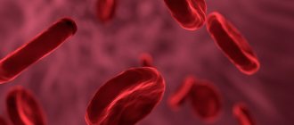
Another component of blood are formed elements (cells). They are represented by erythrocytes - red blood cells, blood leukocytes - white blood cells, platelets - blood platelets. It is red blood cells that answer the question why blood is red.
Red blood cells
At the same time, about 35 billion red blood cells move through the circulatory system. Appearing in the bone marrow, red blood cells form hemoglobin in the blood, which is a red pigment rich in protein and iron. The task of hemoglobin is to deliver oxygen to vital parts of the body and remove carbon dioxide. Red blood cells live on average 4 months, then they disintegrate in the spleen. The process of formation and breakdown of red blood cells is continuous.
Red blood cells give the blood a red color
Why are veins blue and not red?
In fact, of course, although the veins carry dark burgundy blood, unlike the bright scarlet arterial blood, they are not at all blue in color. They are red, like the color of the blood that flows through them. And you shouldn’t believe in the theory that you can find on the Internet that the blood actually runs through the vessels is blue, but when cut and in contact with air it instantly turns red - this is not so. Blood is always red, and why is described above in the article.
The veins only appear blue to us. This is explained by the laws of physics about the reflection of light and our perception. When a beam of light hits the body, the skin reflects some of all the waves and therefore looks light, well, or different, depending on melanin. But it transmits the blue spectrum worse than red. But the vein itself, or rather the blood, absorbs light of all wavelengths (but less, in the red part of the spectrum). That is, it turns out that the skin gives us a blue color for visibility, and the vein itself gives us red. But, interestingly, the vein actually reflects even a little more red than the skin in the blue spectrum of light. But why then do we see veins blue or cyan? And the reason, in fact, lies in our perception - the brain compares the color of the blood vessel against the bright and warm tone of the skin, and in the end shows us blue.
Why don't we see other vessels through which blood flows?
If a blood vessel is located closer than 0.5 mm to the surface of the skin, then it generally absorbs almost all blue light, and reflects much more red light - the skin looks healthy pink (ruddy). If the vessel is much deeper than 0.5 mm, then it is simply not visible, because the light does not reach it. Therefore, it turns out that we see veins that are approximately located at a distance of 0.5 mm from the surface of the skin, and why they are blue has already been described above.
Why can't we see arteries from under the skin?
In fact, about two-thirds of the blood volume is permanently in the veins, which means they are larger than other vessels. In addition, arteries have much thicker walls than veins, because they have to withstand greater pressure, which also prevents them from being sufficiently transparent. But even if the arteries were visible from under the skin as well as some veins, it is assumed that they would have approximately the same color, despite the fact that the blood running through them is brighter.
What color are veins actually?
If you've ever cooked meat, you probably already know the answer to this question. Empty blood vessels are reddish-brown in color. There is not much difference in color between arteries and veins. They differ mainly when viewed in cross section. Arteries are thick-walled and muscular, while veins have thin walls.
Also learn about the main vein diseases worth knowing, as well as about varicose veins and their treatment.
Blue blood
As for aristocrats, the expression “blue bloods” arose due to the paleness of their skin. Until the twentieth century, tanning was not in fashion, and the aristocrats themselves, especially women, hid from the sun, which protected their skin from premature aging and looked appropriate for their status, that is, they differed from the serfs who “plowed” all day in the sun. We now understand that pale skin color with a blue tint is actually a sign of less health.
But scientists also claim that there are about 7,000 people in the world whose blood has a blue tint. They are called kyanetics (from the Latin cyanea - blue). The reason for this is not the same hemoglobin. Their protein contains more copper than iron, which during oxidation acquires a blue tint instead of the red we are accustomed to. These people are considered more resistant to many diseases and even injuries, as their blood is said to clot several times faster and are not susceptible to many infections. In addition, there are different theories about the origin of kianeticians, including that they are descendants of aliens. There is not much information about them on the Internet, but there are articles in foreign publications where the birth of such children is explained by the abuse of rudimentary drugs long before conception. As they say, “Don’t smoke, girl, the children will be green!”, but the results from birth control may turn out blue (meaning the color of blood).
- About company
- Purchase terms
- News
- Articles
- Adviсe
- Contacts
- Pharmacy terms
- Reviews
- Pharmacy Online Savings
Squeezing a pimple and getting sepsis is a reality for people with weakened immune systems. A bacterial infection that causes inflammation of the sebaceous gland can invade healthy tissue and blood. In this case, new ulcers will appear next to the squeezed pimple, and the pimple itself will turn purple and increase in size. If you do nothing, the source of inflammation will expand, covering new areas.
Leukocytes are always on guard
At the final stage, a scar is formed. Epithelization of the restored area is also underway.
Staphylococci - a source of inflammation
The immune system deals quickly with ordinary bacteria. Another thing is pyogenic microorganisms, such as staphylococci, streptococci, meningococcus, gonococcus, Pseudomonas aeruginosa, E. coli. Having the ability to attach to each other, they turn into large objects. As a result, macrophages cannot cope with them. It is also important that pyogenic bacteria have a high division rate.
The presence of abscesses, boils, and carbuncles does not mean a 100% chance of developing sepsis. Generalized inflammation is promoted by immunodeficiency resulting from severe illness, severe blood loss, surgery, poor nutrition, or use of immunosuppressive drugs.
Symptoms of blood poisoning
Regardless of the strain of the bacterial pathogen, the clinical picture of sepsis is the same:
- elevated temperature;
- chills, excessive sweating;
- low blood pressure;
- migraine-like headache;
- arrhythmia (more than 90 heart beats per minute);
- dyspnea;
- weakness,
- lack of hunger;
- nausea, vomiting;
- rash;
- large foci of inflammation in the form of reddened, swollen areas; purulent exudate may flow from the wound.
The conglomeration of symptoms depends on the location of infection and the intensity of development of the bacterial infection.
Therapeutic measures for sepsis
Open purulent wounds are treated by removing exudate and necrotic formations. Affected tissues are regularly treated with antiseptics (levomekol) and antibiotics (amoxicillin). If the inflammation is hidden and is not accessible for surgical treatment, powerful antibacterial drugs are prescribed. Since pyogenic microorganisms quickly adapt to antibiotics, it is necessary to identify their sensitivity to the drugs used.
In addition, measures are taken to strengthen the patient’s immune system. This includes: high-calorie nutrition, the use of autovaccines, blood transfusions, and the use of immunostimulants. Timely elimination of pathogenic factors in the case of sepsis helps the patient get back on his feet.
Blood constantly circulates throughout the body, providing transport of various substances. It consists of plasma and a suspension of various cells (the main ones are erythrocytes, leukocytes and platelets) and moves along a strict route - the system of blood vessels.
Content
Venous blood - what is it?
Venous – blood that returns to the heart and lungs from organs and tissues. It circulates through the pulmonary circulation. The veins through which it flows lie close to the surface of the skin, so the venous pattern is clearly visible.
This is partly due to a number of factors:
- It is thicker, rich in platelets, and if damaged, venous bleeding is easier to stop.
- The pressure in the veins is lower, so if a vessel is damaged, the amount of blood loss is lower.
- Its temperature is higher, so it additionally prevents rapid heat loss through the skin.
The same blood flows in both arteries and veins. But its composition is changing. From the heart it enters the lungs, where it is enriched with oxygen, which it transfers to the internal organs, providing them with nutrition. The veins that carry arterial blood are called arteries. They are more elastic, blood moves through them in spurts.
Arterial and venous blood do not mix in the heart. The first passes along the left side of the heart, the second - along the right. They are mixed only in case of serious heart pathologies, which entails a significant deterioration in well-being.
Lymphatic system
The lymphatic system is formed by lymphatic vessels and lymph nodes located along their course. Lymph circulates in it, similar in composition to blood plasma. Lymph, formed by the absorption of fluid from tissues through lymphatic capillaries, enters the lymphatic vessels, passing through the lymph nodes, where it is filtered, through the lymphatic ducts and trunks it enters directly into the venous system. The lymphatic system, being part of the cardiovascular system, together with the venous network ensures the outflow of water and their waste products from organs and tissues. The permeability of lymphatic vessels is greater than that of capillaries and venules, therefore, through lymph it is possible to remove large particles: colloidal solutions of proteins, fat emulsions, cell breakdown products and microbial bodies.
What is the systemic and pulmonary circulation?
From the left ventricle, the contents are pushed out and enter the pulmonary artery, where they are saturated with oxygen. It is then distributed throughout the body through arteries and capillaries, carrying oxygen and nutrients.
The aorta is the largest artery, which is then divided into superior and inferior. Each of them supplies blood to the upper and lower parts of the body, respectively. Since the arterial system “flows around” absolutely all organs and is supplied to them with the help of a branched system of capillaries, this circle of blood circulation is called large. But the arterial volume is about 1/3 of the total.
Blood flows through the pulmonary circulation, which has given up all the oxygen and “taken away” metabolic products from the organs . It flows through the veins. The pressure in them is lower, the blood flows evenly. It returns through the veins to the heart, from where it is then pumped to the lungs.
General information
For some reason, almost all people are sure that arterial blood is the type that flows in arterial vessels. In fact, this opinion is wrong. Arterial blood is enriched with oxygen, which is why it is also called oxygenated. It moves from the left ventricle to the aorta, then goes through the arteries of the systemic circulation. After the cells are saturated with oxygen, the blood turns into venous and enters the veins of the BC. In a small circle, arterial blood moves through the veins.
You may be interested in: Aromatase inhibitors: purpose and list of drugs
Different types of arteries are located in different places: some are deep in the body, while others allow you to feel the pulsation.
Venous blood moves through the veins into the CD and through the arteries into the MC. There is no oxygen in it. This liquid contains a large amount of carbon dioxide, decay products.
What is venous blood rich in?
When the blood reaches the organs, it gives them oxygen, in return it is saturated with metabolic products and carbon dioxide, and acquires a dark red hue.
A large amount of carbon dioxide is the answer to the question why venous blood is darker than arterial blood and why veins are blue. It also contains nutrients that are absorbed in the digestive tract, hormones and other substances synthesized by the body.
Its saturation and density depend on which vessels the venous blood flows through. The closer to the heart, the thicker it is.
If dark
Venous blood itself is dark in color. This is due to the fact that it flows from the organs, saturated with a large amount of carbon dioxide and giving off oxygen. A rich dark red tint of venous blood is considered normal. However, if the blood has an almost black tint, this indicates serious disorders in the body that need to be treated.
Symptoms and signs
With dark venous blood, a person can not only see a non-standard color of the blood, but also identify disorders based on a number of symptoms, which include:
- tingling in the legs and arms;
- excessive nervousness and anxiety;
- constant yawning and desire to sleep;
- with any damage to the skin, blood flows slowly;
- shortness of breath and rapid heartbeat;
- tinnitus, which is sometimes accompanied by blurred vision or hearing;
Causes
The reason for dark and thick venous blood is the large number of red blood cells it contains. An indicator of this will be hemoglobin. Normally, the hemoglobin level in men should be 115, and in women 125 g/l.
Increased production of red blood cells is associated with disturbances in the functioning of the body, which may be associated with:
- loss of immunity;
- sexually transmitted diseases;
- dehydration;
- pregnancy and difficult childbirth;
- stress;
- great physical activity;
- oncological diseases;
- sudden changes in climate and diet.
Increased production of red blood cells is called erythremia.
Treatment methods
Too dark and thick blood causes serious changes in the body over time, so the treatment of such a deviation must be approached carefully.
Often for complete recovery it is necessary:
- review your nutrition system, drink at least 2 liters every day. water;
- eliminate stressful situations;
- undergo examination and treatment of cancer. And if indicated, special treatment or surgical removal of the tumor;
- undergo drug therapy, which includes the use of drugs such as:
Myelosan
Myelosan is a drug that helps normalize the production of red blood cells.
Helps thin the blood and restore its composition. The drug is prescribed to be taken at a dose of 125-200 mg per day for 30-40 days.
Aspirin
Aspirin is used to improve blood flow. The drug prevents blood clots from platelets from forming inside the flow. Take it 200-300 mg 2-3 times a day for 2-3 weeks.
Chime
Curantil improves the condition of blood vessels and helps restore blood supply. Take the medicine 75 mg 4-5 times a day for 1-2 months.
Herbal teas
Teas infused with medicinal herbs are actively used to thin the blood:
- Chamomile, lemon balm or thyme in the amount of 3 tbsp. l. pour 400 ml of boiling water.
- The infusion is left for half an hour.
- Then the composition is filtered and drunk 200 ml 3-4 times a day instead of tea. The product can be diluted with water or softened with honey.
Meadowsweet
The herb meadowsweet helps increase blood flow.
It improves cerebral circulation and restores blood pressure:
- 1 tsp. herbs must be poured 1 tbsp. boiling water
- The composition is infused under the lid for 20-30 minutes.
- Then drink the decoction ½ tbsp. 3 times a day for 1 month.
Celery and apple
Fresh celery and apple have a unique composition that cleanses the blood and restores its consistency and structure:
- To prepare the product 300 gr. celery should be finely chopped or grated.
- To the mixture you need to add 2-3 peeled apples, which are also finely chopped.
- Then add 2 finely chopped garlic cloves and 2 tbsp. l. parsley
- The mixture is flavored with ½ part lemon and poured over 2-3 tbsp. l. olive oil. The salad should be eaten immediately after preparation and then at intervals of 3 days for 1 month.
Venous blood performs an important function in our body and flows through the veins of the circulatory system, removing all harmful substances. Any disturbance in blood flow can create stagnation or affect the quality of venous blood in the shortest possible time, so the disease should be treated immediately after the appearance of the first symptoms or unfavorable tests.
It is not recommended to self-medicate when such disorders develop, because only a specialist can make a diagnosis, prescribe competent treatment in accordance with the state of health and monitor the dynamics of improvement to fully restore the body’s functioning.
Why are tests taken from a vein?
This is due to the type of blood in the veins - saturated with metabolic products and vital functions of organs. If a person is sick, it contains certain groups of substances, remains of bacteria and other pathogenic cells . In a healthy person, these impurities are not detected. By the nature of the impurities, as well as by the level of concentration of carbon dioxide and other gases, the nature of the pathogenic process can be determined.
The second reason is that venous bleeding when a vessel is punctured is much easier to stop. But there are times when bleeding from a vein does not stop for a long time. This is a sign of hemophilia, a low platelet count. In this case, even a minor injury can be very dangerous for a person.
How to distinguish venous bleeding from arterial bleeding:
- Assess the volume and nature of leaking blood. The venous flows out in a uniform stream, the arterial flows out in portions and even in “fountains”.
- Determine what color the blood is. Bright scarlet indicates arterial bleeding, dark burgundy indicates venous bleeding.
- Arterial is more liquid, venous is thicker.
How to stop venous bleeding?
With minor damage to the veins of the extremities, it is often enough to create an artificial outflow of blood by raising an arm or leg above the level of the heart. A tight bandage should be applied to the wound itself to minimize blood loss.
If the injury is deep, a tourniquet should be placed above the damaged vein to limit the amount of blood flowing to the injury site. In summer you can keep it for about 2 hours, in winter - for an hour, maximum one and a half. During this time, you need to have time to deliver the victim to the hospital. If you hold the tourniquet longer than the specified time, tissue nutrition will be disrupted, which threatens necrosis.
It is advisable to apply ice to the area around the wound. This will help slow down your blood circulation.