Hydrocephalus is characterized by excessive accumulation of cerebrospinal fluid in the natural cavities of the brain. At the Yusupov Hospital, neurologists examine patients with hydrocephalus using modern equipment from leading manufacturers in the USA, Italy, and Germany. After determining the cause and severity of the disease, at a meeting of the expert council with the participation of professors, neurologists of the highest category, and neurosurgeons, a decision is made on the tactics of managing patients with hydrocephalus.
In recent years, to reduce intracranial pressure and restore normal circulation of cerebrospinal fluid, neurosurgeons have widely practiced progressive endoscopic techniques that do not require the installation of a shunt. They are performed by neurosurgeons at partner clinics of the Yusupov Hospital. If hydrocephalus of the brain develops in children, shunting may be the only operation if endoscopic intervention is not possible.
What is hydrocephalus?
The term hydrocephalus is formed from two Greek words: “hydro” - water and “cephalus” - head (“dropsy of the brain”). In hydrocephalus, there is a significant buildup of cerebrospinal fluid (CSF) inside cavities of the brain called ventricles. An increase in the volume of cerebrospinal fluid (CSF) occurs when the balance between the production of cerebrospinal fluid and its absorption and further removal from the body is disturbed. This may be the result of a deficiency in the circulatory system or excessive production of cerebrospinal fluid. Hydrocephalus can be congenital or acquired. The concept of congenital hydrocephalus means that it appears from the moment of birth. The disease can be provoked by various factors: traumatic brain injury, brain tumor, necrosis, meningitis, etc. The frequency of this disease is high throughout the world. According to the literature, hydrocephalus affects 5-15 children out of every thousand newborns. Treatment consists of diverting CSF outside the cerebrospinal fluid system into body cavities where it can be absorbed (absorbed). An effective operation allows a child to live and develop normally, study in a regular school, and an adult to return to a full, active life. However, patients and their loved ones must be able to recognize the signs and symptoms of postoperative complications so that medical care can be provided in a timely manner.
The main types of hydrocephalus
At the initial consultation, the neurosurgeon will listen to the patient’s complaints, but will be able to make an accurate diagnosis only as a result of a comprehensive diagnosis. Special studies make it possible to determine what stage of the disease we are talking about - advanced or mild hydrocephalus.
There are quite a large number of varieties of dropsy:
- by origin, acquired and congenital diseases are distinguished;
- according to the characteristics of the course, hydrocephalus can be closed (occlusive), open (non-occlusive) and mixed;
- by location - internal and external hydrocephalus;
- according to clinical signs - acute, subacute, chronic degrees of hydrocephalus.
The dynamics of pathology development is also important. The most dangerous is considered progressive hydrocephalus, in which the number of symptoms constantly increases. The mild type is considered a regressive disease. With stable hydrocephalus, significant changes in the human body, as a rule, do not occur.
Anatomy and physiology
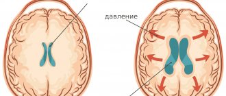
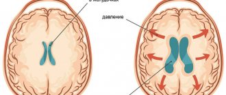
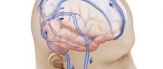
In order to better know and understand the disease, we will give you some information about the anatomy of the skull, the structure of the brain, as well as the process of formation and absorption of cerebrospinal fluid (Fig. 1). The brain occupies most of the cranial cavity. It is penetrated by a large number of blood vessels and is surrounded by cerebrospinal fluid as a buffer. The fluid is located in 4 cavities (ventricles) located inside the brain. The ventricles have delicate structures known as villous plexuses (choroid plexuses). These structures produce a significant amount of CSF - about 500 ml per day. The fluid circulates continuously and contains a large number of substances essential for nutrition and ensuring the normal functioning of the nervous system.
CSF also provides a protective cushion for the brain. CSF circulates in the ventricular system and is removed through 3 holes in the 4th ventricle and then enters the subarachnoid space surrounding the brain and spinal cord.
CSF constantly circulates in the brain and spinal cord, constantly being in the process of a) formation, b) circulation and c) absorption. In a healthy body these processes are balanced. Hydrocephalus develops if CSF is not cleared from the ventricular system through the cerebrospinal fluid tract. Less commonly, hydrocephalus is caused by excess production of CSF, such as with villous plexus papilloma.
Causes
Pathology most often develops in newborns, as well as people of retirement age, although people who have not reached it are also at risk of developing this problem. The maximum chance of signs of hydrocephalus appearing is in those who become ill during fetal development. For every 1000 births there are approximately 1-2 such cases.
Several pathological conditions contribute to an increased volume of cerebrospinal fluid (cerebrospinal fluid):
- disruption of outflow due to the formation of traffic jams in the outflow tract;
- deterioration of fluid absorption by arachnoid granulations;
- too much cerebrospinal fluid production.
So far, all the reasons for the development of pathology have not yet been sufficiently studied. Congenital hydrocephalus is often a consequence of a brain defect, due to which there is a deterioration in the circulation of cerebrospinal fluid for the following reasons:
- hemorrhage into the cerebral ventricles of the fetus;
- incomplete healing of the spinal column;
- intracranial hemorrhage;
- infectious diseases;
- traumatic brain injury;
- congenital genetic pathologies;
- central nervous system tumors;
- arachnoid cysts.
Acquired hydrocephalus can develop in any person due to certain diseases or mechanical injuries:
- intracerebral hemorrhage;
- formation of venous blood clots in the brain;
- stroke;
- meningitis;
- traumatic brain injury;
- development of a tumor in the brain.
Sometimes the vessels through which cerebrospinal fluid flows are narrowed at birth. This pathology can be compensated thanks to the internal reserves of the body. Then hydrocephalus does not appear in the newborn, causing signs of concern only as the child grows older.
In older people, hydrocephalus is called a normotensive disease. This is caused by the situation that the volume of cerebrospinal fluid increases without causing an increase in its pressure. In this condition, intracranial edema occurs, which impairs the functioning of the brain. Conditions in which the natural absorption of cerebrospinal fluid is disrupted increase the risk of developing pathology, although more often the mechanism of development of the disease is unclear. There is a higher risk of infectious meningitis in people who have suffered a traumatic brain injury.
Diagnostics
In newborn children, the bones of the skull have not yet fused and hydrocephalus is determined visually. The head enlarges, the bones of the skull diverge, the fontanelle is tense and bulging; the skin is thin and shiny; The veins in the hairline area look full and swollen. Also symptoms are: vomiting, apathy, excitability, downward displacement of the eyes (“setting sun symptom”), etc. However, when the cranial sutures are not fused, the signs and symptoms of increased intracranial pressure seem subtle.
In older children and adults, the bones of the skull are fused and, when the ventricles expand, compression of the brain tissue occurs. Symptoms of increased intracranial pressure appear: headaches, nausea, vomiting, blurred vision, lack of coordination, psychopathological personality changes, lack of concentration and lethargy. These symptoms require additional instrumental diagnosis.
Diagnosis of hydrocephalus
Signs of the disease are often detected while the fetus is still in the womb during ultrasound. Then the disease is diagnosed by a doctor after the baby is born. The main indicator for determining pathology is the large diameter of the skull. In older children, as in adults, the doctor evaluates the patient’s thinking, his gait, the presence of signs of urinary incontinence, and the condition of the ventricles using MRI images.
To verify the presence of congenital hydrocephalus, CT or MRI are used, which allow:
- assess the state of the brain;
- identify excess cerebrospinal fluid volume;
- detect increased cerebrospinal fluid pressure;
- see structural changes in brain tissue.
Normal pressure hydrocephalus is more difficult to identify because its symptoms are largely similar to neurodegenerative processes. This variant of pathology is diagnosed according to the following signs:
- walking disorder;
- mental disorders;
- urinary incontinence;
- excess volume of cerebrospinal fluid.
With any variant of the disease, it is important to make an accurate diagnosis in a timely manner so as not to be late with surgical intervention that will eliminate the severity of symptoms or reduce their intensity.
To find out how effective the operation will be, some additional measures are necessary:
- Lumbar puncture. A small amount of cerebrospinal fluid is removed through a puncture made in the lower back. The cerebrospinal fluid pressure is then measured. The procedure allows you to slightly reduce it in the cerebral ventricles in order to neutralize severe symptoms.
- Lumbar drainage. It is necessary when the previous procedure was ineffective. A catheter is installed between the lumbar vertebrae, through which the cerebrospinal fluid comes out for several days. All this time the patient is observed in the hospital. Antibiotics are administered to prevent infection.
- Infusion test. This is a stress diagnostic option that helps assess how much the brain is able to absorb cerebrospinal fluid.
- ICP measurement. To perform the procedure, a hole is drilled into the skull bone. A sensor in the form of a fiber-optic cable is installed through it. The event is carried out while the patient is in the hospital for at least 24 hours. The sensor detects surges in intracranial pressure, sending values to a recording device.
When an event involves the need to measure ICP, various sensor options are used:
- Intraventricular catheter. This is the most accurate way to measure. The device is brought to the lateral cerebral ventricle through a hole drilled in the skull bone. The instrument helps to simultaneously ensure the drainage of excess fluid.
- Subdural sensor. The device is attached under the dura mater of the brain. It is used if there is an urgent need to check ICP. The method allows you to quickly identify changes in ICP.
- Epidural sensor. The instrument is attached between the dura mater of the brain and the cranial bone, having previously drilled a hole in it. The procedure is less traumatic than others, but has a significant drawback - excess cerebrospinal fluid cannot be drained during the procedure.
When installing sensors, local anesthesia is required. It is also sometimes necessary to additionally inject the patient with sedative medications in order to reduce anxiety and relax the patient as much as possible.
Typically, sensors used to measure ICP are administered to people in intensive care, often in the most critical condition. The necessary indications for such an event are severe trauma to the skull, pathologies of the brain that cause swelling and depression of consciousness up to a coma. In patients who have undergone brain surgery, such a monitoring sensor helps detect increasing swelling.
Drainage of cerebrospinal fluid also helps to reduce high ICP. It is performed by installing a ventricular catheter, intravenous injection of certain medications, and changing the method of ventilation (in a situation where the patient breathes through a tube inserted into the trachea). Normal ICP is 1–20.
Installing sensors is a risky undertaking, as it can cause the following complications if done incorrectly:
- bleeding;
- irreversible damage in a situation with prohibitive ICP values;
- “herniation” of the brain;
- damage to brain tissue during catheter insertion;
- infectious complications;
- inability to accurately determine the location of the ventricle in order to place a catheter to it;
- other risks present during general anesthesia of the patient.
Types of diagnostic tests
- Ultrasound is a simple, inexpensive test that helps evaluate the extent to which the ventricles of the brain are dilated. Currently, it is the simplest and safest method for diagnosing hydrocephalus.
- Computed tomography (CT) - This is a technique of drawing with a thin beam the contours of the skull, brain, ventricles and subarachnoid space. It is carried out to determine the size and shape of the ventricles and identify abnormalities such as tumors, cysts or other pathologies.
- Magnetic resonance (nuclear magnetic resonance NMR) is a non-surgical diagnostic method that uses radio signals and a magnet. MRI data determines the shape and severity of hydrocephalus. These studies are indispensable for clarifying the causes of dropsy.
- Cisternography (radiography of the cisterns at the base of the skull) is a test that requires the injection of a radioactive substance into the CSF. It is used to clarify the type of hydrocephalus: communicating or obstructive, as well as to determine the direction of CSF flow.
- Pneumoencephalography is now used much less frequently than in the past. In some cases, it is necessary to pump air with a needle into the spinal cord.
- Angiography (X-ray of blood vessels) is a special technique for injecting a contrast agent into the arteries that cross the brain. After some time, anomalies are detected at the level of blood vessels and the presence or absence of pathological disorders.
- A neuropsychological examination consists of a series of questions and answers to identify the presence of abnormalities in the functioning of the brain.
Treatment methods for hydrocephalus in Israel
The treating specialist chooses what treatment to prescribe for hydrocephalus. It primarily depends on the results of the examination. Most often, in adult patients and children, external hydrocephalus of the brain is treated surgically using endoscopic ventriculocisternostomy, neuroendoscopy or shunting.
What is the neuroendoscopy method?
Neuroendoscopy is considered one of the safest operations for the treatment of mixed replacement hydrocephalus of the brain. This method is used more often than bypass surgery. Neuroendoscopy is recommended in cases where the cause of the accumulation of excess fluid is a blockage in its outflow. The purpose of this operation is to create an outflow of fluid from the cerebral ventricle into the spinal canal.
How is the treatment carried out:
A small diameter hole is drilled in the skull, into which a special catheter is installed - endoscopic. A catheter is inserted into the third ventricle of the brain. A small hole is made at the bottom of the ventricle, which, in fact, will ensure the outflow of fluid. The procedure lasts about an hour and is performed under general anesthesia. The advantage of this method is that infection occurs much less frequently than during other operations, such as bypass surgery.
Advantages of neuroendoscopy
During the operation, the doctor makes a small incision. Thus, minimal trauma to the brain is caused and the number of scars is reduced. Postoperative pain is not very severe. The recovery period takes less time than other types of operations. Neuroendoscopy is a complex operation, the successful implementation of which requires not only modern equipment, but also the experience of neurosurgeons. This method is also used to remove brain tumors.
Treatment
Currently, occlusive (obstructive) hydrocephalus is treated surgically. Surgical intervention consists of draining excess cerebrospinal fluid outside the cerebrospinal fluid system: into the abdominal (abdominal) cavity or into the atrium. Sometimes CSF may be drained into the pleural cavity. In these cavities, cerebrospinal fluid is absorbed and excreted along with waste products of the body.
To drain the CSF, the surgeon implants a drainage system (shunt). The system material is silicone and polypropylene. Both of these materials are well tolerated by the body. All elements of the system are implanted under the skin; there are no external areas.
Shunt System Components
The system consists of two catheters and one one-way valve. The ventricular catheter is located in the ventricle of the brain, and the peripheral (peritoneal or cardiac) catheter is placed in the abdominal cavity or in the right atrium, respectively. Both catheters are connected to a valve that regulates the unidirectional flow of cerebrospinal fluid. The valves are designed to operate in different pressure ranges (high, medium, low and very low). The neurosurgeon, having determined the patient’s intracranial pressure, selects the appropriate valve, depending on the severity of the disease, the patient’s age and clinical nuances.
Almost all valve models have a reservoir that your doctor can use to “bleed” the system to determine if it is working properly. From the reservoir, by inserting a thin needle through the skin, you can take samples of cerebrospinal fluid for laboratory tests or administer medications. Patients and their loved ones are not recommended to test the drainage system by “bleeding” the reservoir. This action can be dangerous unless your doctor has given you specific instructions about it. In patients suffering from non-communicating (obstructive) hydrocephalus, cerebrospinal fluid should be drained from the cerebral ventricle using a ventricular catheter. In patients with communicating hydrocephalus, a CSF drainage system is implanted from the lumbar space of the spine into the abdominal cavity - the lumboperitoneal system.
Treatment of hydrocephalus in children
Treatment of hydrocephalus in children
HYDROCEPHALUS IS THE MOST COMMON CAUSE OF SURGICAL INTERVENTIONS ON THE CENTRAL NERVOUS SYSTEM ORGANS IN CHILDREN
Hydrocephalus
, hydrocele - a disease characterized by excessive accumulation of cerebrospinal fluid in the ventricular system of the brain as a result of difficulty in moving it from the place of formation (cerebral ventricles) to the place of absorption into the circulatory system (subarachnoid space) -
occlusive hydrocephalus
, or as a result of malabsorption -
aresorptive hydrocephalus
.
The frequency of occurrence is 1 case in 2000-4000 newborns, more common in boys.
Causes of occurrence.
Hydrocephalus occurs at any age, but most often in young children, due to various reasons: tumors, infectious and inflammatory diseases, traumatic brain injury, congenital anomalies.
Hydrocephalus in a newborn can be caused by a birth craniocerebral injury, infectious diseases suffered by the mother during pregnancy (cytomegalovirus infection), leading to disruption of the ventricular system of the fetal brain. This, in turn, leads to difficulty in the circulation of cerebrospinal fluid and/or its excessive formation. In addition to congenital hydrocephalus, acquired hydrocephalus can also develop (most often in the first months of a newborn’s life) after meningitis, meningoencephalitis, head injuries, intoxication, intraventricular hemorrhages, etc.
Impaired circulation of cerebrospinal fluid (CSF) leads to increased pressure in the cranial cavity and the occurrence of the so-called hypertensive-hydrocephalic syndrome. As a result of the pressure exerted on areas of the brain, vision begins to decrease, convulsions occur, compression of the brain stem is manifested by oculomotor disorders (strabismus, limited upward gaze (the “setting sun” symptom)), weakness in the upper and lower extremities. This can lead to death, severe neurological disorders, and decreased intellectual abilities.
Manifestations
. The most characteristic sign of hydrocephalus in newborns is an accelerated growth of head circumference, leading to a visually well-defined hydrocephalic shape of the skull, greatly increased in volume. Signs of hydrocephalus include a bulging, tense fontanelle, frequent tilting of the head, and a downward displacement of the eyeballs. In places where normal fusion of the skull bones has not occurred, rounded pulsating protrusions may form. Strabismus and nystagmus (involuntary eye movements with high frequency) often occur. There is high excitability due to headaches, the child does not eat well, often cries, vomits, and is lethargic. Sometimes you may notice a decrease in vision and hearing.
Hydrocephalus in older age is characterized by headaches, especially in the morning, nausea and vomiting at the height of headache, dizziness; the size of the head is not increased.
These graphs reflect normal head circumference depending on the age of the child in boys and girls.
Diagnostics.
The most informative are magnetic resonance and computed tomography, which reveal sharply enlarged ventricles of the brain. In young children with an open large fontanelle, neurosonography (ultrasound examination of the brain) is also very informative.
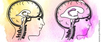
Normal ventricles of the brain.
CT picture of hydrocephalus (dilatation of the lateral ventricles).
IT IS IMPORTANT TO UNDERSTAND THAT THERE IS NO EFFECTIVE DRUG FOR TREATING HYDROCEPHALUS.
MEDICATION TREATMENT OF HYDROCEPHALUS IS MAINLY APPLIED IN THE FORM OF TEMPORARY MEASURES, ESPECIALLY IN CASES WHEN THE DECISION ABOUT SURGICAL TREATMENT HAS NOT BEEN MADE AND MAY BE DELAYED.
ANNUALLY, AT THE NEUROSURGICAL DEPARTMENT OF THE ROSTOV REGIONAL CHILDREN'S CLINICAL HOSPITAL, ABOUT 150 OPERATIONS FOR SURGICAL TREATMENT OF HYDROCEPHALUS ARE CARRIED OUT FOR CHILDREN IN THE ROSTOV REGION AND IN NEIGHBORING REGIONS OF THE SOUTH RUSSIA
Surgical treatment of hydrocephalus in children.
The main method of treating hydrocephalus is endoscopic ventriculostomy of the third ventricle, which consists in creating a connection between the third ventricle and the interpeduncular cistern by dissecting the bottom of the cistern. If endoscopic triventriculostomy is ineffective, a cerebrospinal fluid shunt operation is used - this is a surgical intervention during which a special shunt is installed that drains fluid from the cerebrospinal fluid spaces of the brain into the circulatory system. As a result of the installation of a shunt, fluid does not accumulate in the cranial cavity, and hydrocephalus no longer develops, and a person’s life completely depends on the functioning of this device (shunt).
The entire range of operations performed to treat this pathology is divided into two groups:
1. Operations with drainage of cerebrospinal fluid outside the central nervous system:
-Installation of a ventriculoperitoneal shunt (shunt between the brain and peritoneum);
-Installation of a ventriculoatrial shunt (between the brain and heart);
-Installation of a ventriculovenous shunt (between the brain and veins).
1. “Internal shunting” with the creation of normal channels for the movement of cerebrospinal fluid through the central nervous system systems:
-Torkildsen operation (ventriculocisternostomy). It consists of creating a communication between the lateral ventricle and the occipital cistern by installing a silicone catheter passed under the skin on the back of the head;
-Endoscopic ventriculostomy of the third ventricle. It consists of creating a communication between the third ventricle and the interpeduncular cistern by dissecting the bottom of the cistern in the area of the gray tubercle;
-Implantation of internal stents. It consists of installing stents that expand the Magendie and Luschka holes to normal;
-Plasty of the cerebral aqueduct. It consists of expanding the lumen of the water supply system to ensure normal circulation of cerebrospinal fluid;
-Fenestration of the interventricular septum. It consists of creating an opening between the ventricles through which cerebrospinal fluid can circulate freely.
At the moment, in modern pediatric neurosurgery, only 3 methods of surgical treatment of hydrocephalus are widely used and recognized:
Endoscopic triventriculostomy.
The essence of the method is to create, using a neuroendoscope, a hole in the bottom of the third ventricle for the outflow of fluid into the extracerebral cisterns. As a result, the cerebrospinal fluid flows freely and the person is cured, without shunt dependence. The neuroendoscope is a complex optical system of Hopkins lenses, an illumination channel, a video channel, as well as channels for inlet and outlet of fluid, built into a tube with a diameter of only 6 mm. The operation is performed through a small hole in the skull. The image from the endoscope camera is transmitted to the screen and the doctor sees where the instrument needs to be inserted to restore the outflow of fluid. The method is less traumatic and reliable. Unlike bypass surgery, which takes about an hour, the operation takes only 10-20 minutes.
TO CARRY OUT TRIVENTRICULOSTOMY, THE NEUROSURGICAL OPERATING ROOM OF THE CSTO ROOM IS EQUIPPED WITH MODERN ENDOSCOPIC EQUIPMENT FROM THE LEADING MANUFACTURER
KARL STORZ.
This is what the perforated bottom of the 3rd ventricle looks like.
Endoscopic ventriculostomy of the third ventricle is an effective method for the treatment of obstructive triventricular hydrocephalus; with the correct choice of indications, it allows achieving a lasting cure in 90% of such patients and, therefore, is the preferred method. The risk of serious complications after endoscopic ventriculostomy of the third ventricle is low and amounts to 6.6%. The key to the safety of the operation is: its planning on the basis of complete and high-quality clinical and radiological data and strict adherence to the methodological standard of the operation at all its stages, which together makes it possible to reduce the risk of serious complications by three times. The peculiarity of this surgical intervention is that it is highly effective only for obstructive forms of hydrocephalus.
Ventriculoperitoneal and ventriculoatrial shunting.
Ventriculoperitoneal shunt surgery has been in practice for more than fifty years, being the main standard method for getting rid of almost any form of hydrocephalus. Regardless of the technical nuances of the operation, the final result of VPS is the creation of an artificial outflow of cerebrospinal fluid from the dilated ventricles into the abdominal cavity. In some cases, in order to reduce the risk of trauma to the abdominal organs, in the presence of adhesions after previous abdominal operations, it is possible to use laparoscopic techniques. The shunt system consists of 3 main elements:
— ventricular catheter
, which is installed into the ventricular system through a small burr hole at one of the standard points;
— shunt pumps
regulating the outflow of cerebrospinal fluid from the ventricles; All models of pumps equipped with valves can be divided into three types depending on their throughput, or, in other words, on the level of cerebrospinal fluid pressure (low, medium and high pressure). Shunt systems with valves of a given pressure pose a certain complexity for the surgeon, since an error in selecting the parameters of a shunt system can lead to both excessive and insufficient levels of cerebrospinal fluid drainage; this problem can be solved by shunt systems that have appeared not so long ago, equipped with programmable valves, the “highlight” of a programmable valve is that it is equipped with a device designed for the possibility of remotely changing the level of cerebrospinal fluid drainage;
— peritoneal or atrial catheter
, immersed in the abdominal cavity or atrium to ensure the outflow of cerebrospinal fluid.
This is what a shunt system looks like, consisting of a ventricular catheter, a shunt pump and a peritoneal/atrial catheter
ü IN THE NEUROSURGICAL DEPARTMENT OF GBU RO "CSCH", FOR SURGICAL CORRECTION OF HYDROCEPHALUS ONLY MODERN SHUNT SYSTEMS FROM LEADING FOREIGN MANUFACTURERS, SUCH AS
INTEGRA, AESCULAP, ETC.
Complications.
The key point in the development of complications after liquor shunt operations using shunt systems is the presence of a foreign shunt system in the child’s body. According to various authors, in children in the first year of life, the risk of developing dysfunction of the shunt system (that is, its failure), even with a favorable course, is up to 50% and subsequently decreases by 10% with each subsequent year of the child’s life. The essence of the dysfunction is the impossibility of ensuring the shunt system of normal outflow of cerebrospinal fluid for one reason or another, due to the failure of the main elements of the shunt system, in addition to purely technical issues associated with the breakdown of the valve apparatus of the pump, frequent causes of shunt dysfunction are the development of shunt infection, the formation of pseudoperitoneal cysts in the abdominal cavity (a limited cavity that does not allow cerebrospinal fluid to be normally absorbed into the peritoneum). Clinically, the development of dysfunction is manifested by the development of general cerebral (the child becomes lethargic, adynamic, refuses to eat, nausea, vomiting appears) and focal neurological symptoms (Graefe’s “setting sun” symptom, meningeal symptoms, etc.).
It is important to remember that dysfunction of the shunt system is a serious complication in which repeated surgical intervention is indicated to eliminate it; If there is a suspicion of dysfunction of the shunt system, it is necessary to urgently seek specialized neurosurgical help to examine the child and decide on further treatment tactics!!!
ü Neurosurgical Department of the State Budgetary Institution of the RO “CSTO” operates around the clock and has the ability to conduct emergency surgery for hydrocephalus, including in cases of the development of displacement of shunting systems
Unfortunately, even a successfully performed operation is not a guarantee of hydrocephalus cured for life, since the anatomical dimensions of organs can change, the head can grow (especially in children), the holes in the shunt systems become clogged, etc. Children who have undergone such operations need to be constantly monitored by a neurologist and neurosurgeon in order to promptly identify emerging disorders that require correction. Thus, due to changes in the position of organs or the growth of the head, repeated operations have to be performed to replace the shunt with a more suitable one in terms of parameters.
Surgery and hospitalization
Implantation of the shunt system is carried out by a neurosurgeon under sterile operating room conditions. In preparation for surgery, the hair is shaved to achieve maximum cleanliness. The operation is performed under general anesthesia, usually not for a very long time. The neurosurgeon drills a small hole in the skull bone, then makes a small incision in the dura mater that protects the brain and inserts a ventricular catheter into the lateral ventricle. To implant the rest of the bypass system, another incision is made and a tunnel is created subcutaneously to implant a peritoneal or cardiac catheter. The end of the catheter is carefully inserted either into the abdominal cavity or into the jugular vein leading to the ventricle of the heart. Catheters are connected to the valve using connectors on the valve itself. All parts of the shunt are thus connected into a single drainage system. At the end of the operation, small sterile dressings are applied to each incision.
Immediately after the operation, the patient is taken to the postoperative department. The patient is here from 1 hour to 1 day under close observation, then he is transferred to the ward. Most patients are discharged 7-10 days after clinical rehabilitation.
Diagnosis and treatment methods for dropsy
Diagnosis of hydrocephalus is carried out in several ways. For example, using multislice computed tomography. As a result, the location of the pathology and its nature are determined - closed or open. The second common research method is MRI (magnetic resonance imaging), which provides accurate data on the condition of the brain vessels, the presence of structural changes and inflammatory processes.
In the early stages of identifying hydrocephalus, conservative treatment is prescribed - for example, medication. If the results are unfavorable, surgery is indicated:
- shunting for hydrocephalus involves draining excess fluid through a tube;
- external ventricular drainage - organization of a special drainage that is inserted into the cavity with cerebrospinal fluid;
- surgical removal of a septum, tumor, or blood clot that obstructs the movement of fluid.
Contraindications to surgery to eliminate symmetrical or asymmetrical hydrocephalus are severe cardiac pathologies, inflammatory processes in the nervous tissue, and serious problems with the blood vessels or respiratory system.
Neurosurgeons of the city clinical hospital named after. A.K. Eramishantsev will help cope with problems such as replacement, normotensive, triventricular, vicarious hydrocephalus. Sign up for a consultation to get more accurate answers to your questions. With timely intervention, the rehabilitation period after surgery occurs with minimal discomfort, and within a few days after surgery, adult patients can return to work, and children can return to school and a full-fledged active lifestyle.
Participation in care
Children with implanted shunts should be monitored by a neurosurgeon throughout their lives. Most patients suffering from hydrocephalus, after implantation of a shunt system, can lead a normal life, but constant monitoring by loved ones in collaboration with a neurosurgeon is necessary.
The neurosurgeon monitors each patient to prevent drainage failure. The first time after implantation or re-operation - regularly, with a gradual transition to examinations once a year.
Relatives are advised to be able to recognize early signs of complications. Quick and accurate assessment of health problems is very important. Flu symptoms may mask symptoms of a blocked shunt. Quickly identifying them will allow you to plan a repeat operation and avoid an emergency.
Patients and their loved ones should pay close attention to signs and symptoms of complications. The main causes are: blockage, infection and excessive drainage.
What is needed for the initial diagnosis of hydrocephalus in Israel
To confirm the diagnosis, it is necessary to undergo a primary diagnosis. To do this you need:
- Blood test – $280
- Computed tomography – $630
- Magnetic resonance imaging (MRI) – $1350
- Consultation with a neurosurgeon based on examination results – from $660
To get tested, you can sign up:
- Call us at: +972(77)4450-480.
- By leaving a request on the website.
We will contact you quickly.
Blockage
The main type of complication is blockage of the system. Blockages can occur at any level of drainage. The holes in the ventricular catheter may become clogged with brain tissue or villous plexus tissue. It can also be blocked due to excessive contraction of the ventricular cavity due to too intense drainage of fluid from the ventricular cavity (constricted ventricle syndrome). Intestinal loops or necrotic tissue may adhere to the peritoneal catheter. Shunts inserted into a ventricle of the heart can become blocked by blood clots, pieces of brain matter, or tumor cells.
The shunt system may also be disconnected by disconnection of various elements of the shunt or by changes in the position of the catheter caused by the growth of the child. X-rays are necessary to check the integrity of the shunt.
Partial blockage of the system parts provokes the appearance of symptoms of increased intracranial pressure. Episodic headaches, nausea, apathy and decreased sensory functions may occur. Decreased performance at school or at work is the most common phenomenon in these conditions.
In case of complete blockage, symptoms develop more quickly (headaches, nausea, vomiting, blurred vision, loss of coordination and confusion). The patient falls into a stupor or coma. In such cases, urgent hospitalization is necessary for observation and appropriate treatment.
The surgeon performs a series of tests to locate and determine the extent of drainage blockage. Sometimes it is necessary to remove and replace part or all of the system.
Life after hydrocephalus shunt
The shunt remains in the patient's body for life. Brain shunt surgery has some disadvantages and side effects, but it is still widely used for hydrocephalus to treat and prevent the effects of increased intracranial pressure. The quality of life of patients after hydrocephalus shunting is significantly improved.
Neurosurgeons revise the shunt from time to time. This procedure is performed due to the child’s growth or disruption of the functioning of the shunt - its wear and tear, blockage. Modern shunts are designed for a ten-year service life. It is almost impossible to predict when a shunt will require replacement. Parents whose children have hydrocephalus and require shunting receive detailed instructions from the neurosurgeon on caring for the baby, signs of disruption of the normal operation of the shunt, and safety measures. If the shunt is blocked and the outflow of cerebrospinal fluid is disrupted, neurosurgeons provide patients with emergency qualified medical care.
Infection
Infection is the second type of complication. It poses a significant risk for any surgical procedure, most often when implanting a foreign body.
It manifests itself in the form of redness or suppuration along the edges of the suture or along the path of the drainage system under the skin. The surgeon fixes his attention on these signs. If left untreated, the wound may erode or open, and in more serious cases, the infection may cause chills and fever. Typically, the drain needs to be removed. Sometimes antibiotic therapy can be administered without removing the system.
Since the shunt is a foreign body, the patient may experience an allergic or inflammatory reaction. Inflammation at one of the drainage sites should be immediately shown to a neurosurgeon.
Excessive drainage
Excessive drainage of cerebrospinal fluid occurs when the valve is incorrectly selected according to the pressure parameter. If the valve opening pressure is too low, it can cause excessive drainage, causing the cerebral ventricle to compress and deforming the brain tissue. The patient experiences headaches that are most severe when standing.
In addition, nausea, vomiting, drowsiness and nervous system disorders, in particular double vision, appear. School-age children experience a decline in mental abilities.
Symptoms of hydrocephalus
Hydrocephalus manifests itself differently in each person. Main symptoms:
- Infants may have a large skull, a convex or tense fontanel.
- Older children suffer from constant headaches and blurred/double vision. There are cases when a child becomes completely blind. Children may experience unclear gait and frequent but involuntary urination.
- In adults, there is a decrease in muscle tone. They begin to complain of irritability and drowsiness. Hydrocephalus is often accompanied by vomiting.
Symptoms, as well as treatment for replacement hydrocephalus of the brain, differ depending on age.
More information about shunt systems for the treatment of hydrocephalus
The result of the operation largely depends on the quality of the shunt system. In recent years, in many hospitals, neurosurgeons have been using shunt systems produced by the largest American company Medtronic. The company's engineers, together with leading US neurosurgeons, have developed a range of valves of various models and sizes (including valves for newborns).
One of the latest achievements in the production of liquor drainage devices is the Delta valve. This is a unique valve, has no analogues from other manufacturers. It was created to avoid such a common complication as excessive CSF drainage. If all other valves pass as much fluid as they are designed for pressure, then the Delta valve allows as much fluid as is needed to remain in the ventricle to maintain intracranial pressure within physiological limits. When implanting the Delta valve, the patient is maintained at normal pressure, regardless of the rate of cerebrospinal fluid production and, most importantly, regardless of the patient’s body position (lying/standing).
The technological features of the materials from which the system valves are made prevent deformation and sticking during pumping; the dome of the valve reservoir is designed to withstand repeated punctures with a thin needle (the holes self-tighten). Catheters are made of high-quality latex-free silicone, so they do not stick together or form loops, which significantly reduces the risk of system blockage.
The valves are equipped with connectors for connection to catheters; their design facilitates connection and reduces the possibility of disconnection and disconnection of the system.
A radiopaque mark is applied along the length of the catheters, this allows you to see the shunt on an x-ray. The same substance is applied to the valve with a dot code indicating the pressure of the valve. There are no metal parts in the shunt system. This is very important when conducting CT and NMR studies, because metal will produce artifacts, and a magnet in NMR may shift the location of the system (if it had metal parts).
All systems are sterile and supplied in double sterile packaging. To reduce the risk of infection, Medtronic has developed a unique BioGlide hydrogel. It is applied to the inner and outer surfaces of catheters, as well as the outer surface of the valve, and does not peel off. Before implantation, the neurosurgeon can treat the system parts with antibiotics, and the hydrogel will hold them for 3 days for postoperative antibiotic therapy inside the patient’s body. This way, the risk of infection is minimized.
Discussion
The concept of CHIC is inextricably linked with such pathological conditions as CDV and slit ventricle syndrome (SVS). Understanding the nature of these disorders is key in choosing adequate treatment for this group of patients.
It is necessary to distinguish between simply anatomical reduction in the size of the ventricles, which does not require treatment, with the thyroid gland itself, which has characteristic clinical manifestations. On neuroimaging, narrow or slit-like ventricles are found in 20-53% of children with shunts, while clinically thyroid gland is observed in 1-37% of cases [4, 8-15]. The syndrome develops predominantly in children operated on during the 1st year of life [16]. The average age of manifestation of thyroid gland is 6-7 years [9, 13], although an earlier development of this condition cannot be ruled out [6]. Explaining the causes of the pathology causes a lot of disagreement, and the choice of tactics for thyroid gland remains a subject of debate to this day [1, 17—22]. However, it is quite obvious that all attempts to elucidate the mechanism of development of PTS against the background of CHIC inevitably lead us to the problem of CCD.
CCD refers to a condition where the volume of the growing brain exceeds the available intracranial space. In this case, normal physiological fluctuations in intracranial contents (the volume of intracranial blood and cerebrospinal fluid) lead to an increase in ICP with characteristic clinical manifestations. Various terms are used to describe this pathology in the literature: cephalocranial disproportion [23-26], post-shunt craniosynostosis [4], cranial hyperostosis [27], secondary craniosynostosis [28, 29], shunt-induced craniostenosis [17, 30], secondary microcrania [24] and others.
A. Sandler et al. [31] distinguishes two categories of CDCs:
1) primary CDD - caused by a primary pathological process leading to diffuse thickening of the bones of the arch or premature ossification and closure of the sutures. A striking example of such CCD is primary craniostenosis;
2) secondary CDD is an iatrogenic phenomenon that is initiated by implantation of a shunt system in early childhood.
The incidence of secondary CCD is unknown, since no similar studies have been conducted. Severe, disabling headaches occur in 42% of adolescents with shunts [32]. In a study of 3100 patients undergoing LSO, craniostenosis was detected in 1.6% of patients [1]. In our group of 59 children, 27 (46%) were diagnosed with premature suture closure, while signs of CD were noted in only 3 (5%) of them. It is possible that the approximate incidence of CCD can be indirectly estimated from the incidence of slit ventricle syndrome. It is conceivable that a significant proportion of patients with this syndrome suffer from CCD and their narrow, slit-like ventricles may be a manifestation of a crowded skull.
In CCD, the brain's ability to compensate for physiological fluctuations in intracranial volume is compromised for a number of reasons [33]. First, the discrepancy between normal brain volume and small cranial volume leads to increased extraparenchymal resistance. Secondly, the reduced volume of the ventricles and subarachnoid spaces, which normally participate in compensating for increased ICP, significantly reduces the potential for damping ICP fluctuations [34]. Thirdly, impaired venous outflow during CDD leads to increased pressure in the superior sagittal sinus, which impairs the absorption of cerebrospinal fluid [14]. In addition, venous congestion leads to changes in the turgor of brain tissue, increasing its rigidity and reducing compliance [11, 14].
Clinical manifestations
Typically, such patients are operated on for hydrocephalus at an early age (before 1 year) and, as a rule, they have a history of repeated revisions and multiple visits for chronic severe headaches, despite a functioning shunt. If drug treatment has an effect, it is most often insignificant or short-lived. The severity of the headache limits daily activities, including school or work activities [35, 36]. In contrast to the “low pressure” headaches that occur in a hyperdrainage state, in CCD, lying down does not reduce the severity of pain. Other manifestations of increased ICP may include diplopia, ataxia, dizziness, and disturbances of consciousness. Cases of acute deterioration leading to death have also been described [26].
Diagnostics
In a number of patients, upon examination, a characteristic deformation of the skull is revealed: microcephaly, scaphocephaly, plagiocephaly. X-rays reveal thickening of the bones of the arch, pronounced “finger impressions”, erosion of the internal plate from the pressure of adjacent grooves, thickening or compaction of the diploe, overlapping of bones in the suture area and their synostosis. Neuroimaging demonstrates normal or slit-like ventricles that do not always dilate even during an exacerbation. This is due not only to a decrease in brain compliance, but also to the absence of intraventricular obstruction [22]. On K.T. and MRI reveals a narrowing of the convexital subarachnoid spaces, as well as overfilling of the cranial cavity, especially in the posterior cranial fossa. In this case, both ascending and descending dislocation of the cerebellum can develop [24, 37]. An increase in ICP does not always lead to changes in the fundus [22]. With invasive ICP monitoring in such patients, A. Sandler et al. [31] found a correlation between headaches and the appearance of plateau waves of increased ICP.
Treatment
In case of radiographically confirmed secondary craniosynostosis and the presence of signs of CD (clinical manifestations of intracranial hypertension, narrowing of the subarachnoid spaces and cerebral ventricles), surgical intervention aimed at decompression of intracranial structures is indicated. Additional criteria may include thickening of the bones of the calvarium, pronounced “finger impressions,” and acquired Chiari malformation.
In 1974, F. Epstein et al. [20] published the results of subtemporal craniectomy performed on 2 children with narrow ventricles who had previously received shunt systems. Essentially, this method creates an artificial surgical fontanel, providing additional space for the brain, and also making it possible to palpate the increased pressure. Such decompression can be performed on one or both sides [22]. However, the authors themselves noted the low effectiveness of such interventions and unacceptable cosmetic consequences: due to the protrusion of the temporal flaps, the head took on the shape of a cloverleaf [22]. Another method used by F. Epstein et al. [11] in 2 children with shunts, there was skull fragmentation - “a radical expansion of the skull... by fragmentation of the skull from the coronal suture to the transverse sinus posteriorly and to the squamosal sutures laterally.”
Open reconstructive interventions aimed at increasing the volume of the skull have become widespread [1, 4, 6, 17, 38]. Particularly noteworthy is the fact that in case of secondary dislocation of the cerebellum, supratentorial reconstruction of the skull is pathophysiologically more justified than decompression of the posterior cranial fossa [24]. When performing reconstruction, it is advisable to replace the valve of the shunt system with a valve with a higher pressure or a programmable valve, or to add an anti-siphon device to the system [1].
Another effective method for correcting secondary craniosynostosis and CD is the use of distraction devices. Distraction osteogenesis can be defined as bone growth surgically induced by applying distending forces at the osteotomy site [39]. The simultaneous use of invasive ICP monitoring sensors and gradual distraction of the skull bones makes it possible to individualize the technique in relation to each patient, performing distraction to the required amount, at which ICP parameters are normalized [31].
Emotional support
The physical side of hydrocephalus is only part of the problem of this disease. The patient and his relatives need to take into account his emotional factors.
Although surgery should resolve your hydrocephalus to some extent, you may feel fearful, depressed, irritable, or doubtful. If the patient is a child, it should be taken into account that he has the same feelings as an adult. If your child is feeling out of shape or uncomfortable because they need to visit the doctor frequently or have repeated tests, it is best to reassure them with simple explanations. If he knows what awaits him, he will be more willing to cooperate with you. Children, like adults, as a rule, do not like unpleasant surprises. A calm atmosphere among loving loved ones is the best environment for children. It is advisable to explain the phenomenon of hydrocephalus in words that the child can understand.
It is very important to know how the child feels and be able to explain to him what he is experiencing. The needles hurt. It is natural to cry and want to get rid of them. Being admitted to hospital is a new challenge for your child. You must tell him the truth in order to gain and secure his trust. Sincerity is the best way to maintain your child's trust.
Children over ten years of age are generally able to comprehend more complex concepts. They may associate signs and symptoms with their illness. Restrictions associated with the disease are easier for them to bear. Tell your doctor about your feelings and give him the right to guide you. Some people share their feelings with close friends, others need professional help. The health professionals treating you or your child are interested in your well-being and their goal is to do what is best for you and your loved ones.
Patients and the patient's parents should communicate with their doctor quite frequently. It is important to actively participate in this communication so that your doctor can better understand your needs and the needs of your loved ones.