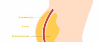
Causes of the disease
As already noted, chronic rheumatic heart disease is an outcome of ARF.
Acute rheumatic fever, in turn, occurs after illnesses such as:
- pharyngitis;
- angina;
- caries.
If the infection develops into a chronic one because it remains untreated, ARF develops. In this case, the human immune system actively produces antibodies that have a damaging effect not only on streptococcus, but also on the heart.
Symptoms and signs of rheumatism
The disease can be suspected by the following manifestations:
- development of the disease 1-2 weeks after tonsillitis, pharyngitis, scarlet fever, erysipelas;
- a sharp increase in body temperature, general weakness;
- joint pain (knee, ankle, elbow, wrist);
- swelling and redness of the skin over the joints, stiffness of movement;
- pain in the heart area;
- shortness of breath, dizziness, headache;
- muscle weakness, involuntary twitching of the limbs;
- pink rash in the form of a ring on the body, passes quickly;
- rheumatic nodules - painless, dense small formations under the skin in the joint area;
- nagging pain in the lower back, darkening, redness of urine.
If such symptoms appear, you should make an appointment with a physician, cardiologist or rheumatologist.
Types of pathology
Rheumatic heart disease affects not only the valve apparatus, but also provokes myocarditis, endocarditis or pericarditis.
Emerging heart defects are classified as follows:
- Defects with stenosis. This pathology is characterized by a narrowing of the valve outlet.
- Vices with deficiency. This pathology is characterized by incomplete closure of the valve leaflets when closing.
- Combined defects. They are characterized by insufficiency and stenosis of the aortic valve.
- Combined defects. This pathology is characterized by mitral regurgitation and aortic stenosis.
Classification of rheumatism
Based on the nature of the flow, the following forms are distinguished:
- acute (up to 3 months);
- subacute (3-6 months);
- prolonged (more than 6 months);
- latent (hidden) - occurs without characteristic symptoms, without laboratory changes, is detected after the formation of heart defects;
- recurrent - wave-like course with rapid development of internal organ failure.
There are active and inactive phases of the disease. In the active, a specific laboratory picture is observed (increased levels of C-reactive protein (CRP) - reflecting acute inflammatory processes, antistreptolysin).
Clinical forms of rheumatism:
- rheumatic carditis - inflammation of the heart tissue;
- polyarthritis ー multiple joint damage;
- ring-shaped erythema - a specific rash on the skin;
- chorea - severe neurological symptoms (hand tremors, muscle weakness, involuntary movements);
- subcutaneous nodules - with the formation of dense nodules under the skin in the joint area.
Symptoms of rheumatic heart disease
Symptoms of pathology can be very different.
Typically patients complain of:
- General malaise, weakness and increased fatigue.
- Shortness of breath not only during exertion, but also at complete rest.
- Cardiopalmus.
- Chest pain during exercise.
- Attacks of lack of air mainly at night, during sleep.
- Sensation of pulsation inside the chest.
- Pulsation of neck vessels.
- Dizziness.
Against the background of heart disease, the following may occur:
- symmetrical swelling of the legs and ankles;
- fainting.
When examining the patient, cyanosis of the nasolabial triangle and blush on the cheeks can be detected. With mitral stenosis in children, developmental delays occur. Patients often complain of weight loss, as well as coughing with sputum (with lesions of the mitral valve). Each symptom is not specific. Therefore, it is very important to constantly monitor the patient’s condition and conduct a comprehensive examination.
Pathological complications deserve special attention.
After a fever, mitral valve insufficiency often develops. It occurs in patients in 60-70% of cases. Isolated aortic valve insufficiency also occurs. Isolated stenosis is the least common. All these pathologies are dangerous to the life and health of the patient.
Causes of acquired heart defects
In most cases, the defects are caused by rheumatic diseases, in particular rheumatic endocarditis (about 75% of cases). The cause may also be the development of atherosclerosis, systemic connective tissue diseases, trauma, sepsis, infections, overload, and autoimmune reactions. These pathological conditions cause disturbances in the structure of the heart valves.
Classification of acquired heart defects
The human heart has four chambers: the left and right atria and ventricles, between which are the heart valves. The aorta emerges from the left ventricle, and the pulmonary artery emerges from the right ventricle.
Between the left heart chambers is a bicuspid valve, the mitral valve. And between the right sections there is a tricuspid valve, another name is tricuspid. In front of the aorta there is the aortic valve, in front of the pulmonary artery there is another valve, the pulmonary valve.
The performance of the heart muscle depends on the functioning of the valves, which, when the heart muscle contracts, allow blood to flow into the next section without obstruction, and when the heart muscle relaxes, do not allow blood to flow back. If the function of the valves is impaired, the function of the heart is also impaired.
Based on the reasons for their formation, defects are classified as follows:
- degenerative, or atherosclerotic, they occur in 5.7% of cases; More often, these processes develop after forty to fifty years, calcium is deposited on the leaflets of the damaged valves, which leads to the progression of the defect;
- rheumatic, formed against the background of rheumatic diseases (in 80% of cases);
- defects that arise as a result of inflammation of the inner lining of the heart (endocarditis);
- syphilitic (in 5% of cases).
Based on the type of functional pathology, defects are divided into the following types:
- simple – valve insufficiency or stenosis;
- combined – insufficiency or narrowing of two or more valves;
- combined - both pathologies on one valve (stenosis and insufficiency).
Depending on the location, the following pathologies are distinguished:
- mitral valve disease;
- tricuspid defect;
- aortic defect.
Hemodynamics can be impaired to varying degrees:
- insignificant;
- moderately;
- expressed.
The mitral valve is affected more often than the aortic valve. Pathologies of the tricuspid valve and pulmonary valve are less common.
Diagnostics
To identify pathology, a specialist:
- Evaluates patient complaints, studies anamnesis, paying special attention to past infectious diseases.
- Conducts a full inspection.
- Orders an ECG. This examination can detect arrhythmia.
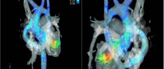
- Prescribes echocardiography. This examination helps to detect defects, determine their type, as well as the presence of circulatory disorders and their severity.
Additionally, the cardiologist can refer the patient to a rheumatologist and cardiac surgeon. Comprehensive diagnostics allows you to quickly make an accurate diagnosis and begin therapy.
Important! Diagnosis is also carried out during therapy. Examinations make it possible to identify the causes of pathology, associated disorders, change the dosage of medications taken and solve other important problems.
Treatment
Pathology therapy is carried out:
- Medication.
- Surgically.
Drug therapy is aimed at relieving signs of heart failure, arrhythmia and preventing complications.
Doctors prescribe the following drugs:
- Beta blockers. These drugs can reduce the manifestations of heart failure and arrhythmias.
- Diuretics. These drugs are prescribed to reduce the load on the heart muscle.
- Anticoagulants and antiplatelet agents. These drugs help prevent thrombotic complications of the pathology.
- Antibiotics. These drugs help prevent the development of bacterial endocarditis.
If the disease is actively developing and threatens serious complications, surgical interventions are performed. One of the popular ones is heart valve replacement.
Indications for surgery are:
- severe chest pain;
- dyspnea;
- frequent fainting;
- chronic heart failure in a severe stage;
- endocarditis.
The intervention is quite complex and lengthy. After surgery, patients have to stay in the hospital for a long time (up to a month).
Today, open-heart interventions are being successfully replaced by minimally invasive ones. They are carried out through a mini-access without cutting the sternum. The intervention is endovascular. It is used in aortic valve replacement. The operation is performed by introducing a prosthesis through the femoral vein. Of course, doctors strive to minimize all consequences and risks. Nevertheless, preference (if possible) is almost always given to drug therapy.
Are you interested in prices for services in our clinic in Moscow? We adhere to a loyal pricing policy. Thanks to this, you do not overpay even for extensive examinations and long courses of therapy. The price of a consultation with a cardiologist is also relatively low. At the same time, you can always count on receiving comprehensive professional support even in advanced situations.
Sign up for treatment of rheumatism at the Kutuzovsky Children's Center
Relying on traditional methods of treatment and letting the disease take its course is categorically not recommended. Rheumatism is a serious disease that affects almost the entire body and is fraught with dangerous consequences. It must be treated under the supervision of a doctor, achieving a transition into stable remission.
A rheumatologist may recommend:
- Taking medications that will relieve inflammation and pain, as well as eliminate infection.
- Physiotherapy. During the period of exacerbation, UV irradiation is indicated, and during remission it makes sense to take a course of UHF, paraffin baths or electrophoresis for prevention.
- Surgery. This option is not excluded, but is rarely used, only in the most severe cases.
To select effective therapy, it is necessary to accurately determine the characteristics of the disease. This requires research. Most often, in addition to laboratory tests, MRI, ultrasound, goniometry and thermometry are used. For patients suffering from rheumatism, movements are difficult and cause pain. Therefore, the Kutuzovsky Treatment and Diagnostic Center made sure that you undergo the examination with maximum comfort.
You can do all the necessary tests on the territory of the medical center. Consultation with a rheumatologist and diagnostic tests are carried out by appointment. Our patients do not have to stand in line or wait long for test results. Come to the diagnostic and treatment center to undergo diagnostics and choose the optimal treatment regimen.
The contents of this article have been checked and confirmed for compliance with medical standards by rheumatologist Larisa Ivanovna Mazanova.
Prevention
The consequences of the pathology are dangerous to the patient’s health. Thrombosis, heart failure, arrhythmia are negative conditions that can cause death. Therefore, doctors pay special attention to preventive measures.
Prevention is divided into:
- Primary.
- Secondary.
Primary measures are aimed at preventing the development of pathology and consist of:
- Proper nutrition.
- Active recreation in nature and hardening.
- Elimination of all existing foci of chronic infections.
- Adequate treatment of pharyngitis, sore throat and other pathological processes.
- Maintaining bed rest.
Rheumatic attacks can be prevented by monitoring with a cardiologist and preventing the formation of bacterial endocarditis.
Secondary preventive measures are aimed at preventing the occurrence of relapses of rheumatic attacks and the progression of pathology.
They consist of periodic administration of antibiotics for 5-7 years after acute rheumatic fever. Also, antibiotics are necessarily prescribed after any (even the smallest) surgical interventions (tooth extraction, tonsils, etc.). It is important to understand that even a diagnosis of CRHD is not a death sentence. With proper therapy and comprehensive preventive measures, it is possible to improve the quality of life and increase its duration even in the presence of heart defects.
YOU can call us: 8 (8452) 98-84-68 and +7-967-500-8468 or
Rheumatism is a disease that develops unnoticed and gradually. It occurs after a streptococcal infection and consists of inflammation of the connective tissue, which is found in all organs and systems of the body. The heart, blood vessels and joints are primarily involved.
In modern medical literature, this term has been replaced by the generally accepted throughout the world “ acute rheumatic fever ”, which is due to the contradictory understanding of the term “rheumatism” in Russia.
Causes
The disease is triggered by special bacteria - group A beta-hemolytic streptococci. Once inside our body, they can cause tonsillitis (tonsillitis), pharyngitis, and lymphadenitis. However, rheumatism can be a consequence of this infection only if the person has certain defects in the immune system. According to statistics, only 0.3-3% of people who have had an acute streptococcal infection develop rheumatism.
Etiology . Infection with β-hemolytic streptococcus of group A. The presence of foci of infection in the nasopharynx (angina, chronic pharyngitis, chronic tonsillitis). Scarlet fever. Genetic predisposition.
Risk factors for developing rheumatism:
- the presence of rheumatism or systemic connective tissue diseases in first-degree relatives (mother, father, brothers, sisters);
- female;
- age 7 - 15 years;
- previous acute streptococcal infection and frequent nasopharyngeal infections;
- content in the body of a special protein - B-cell marker D8/17
What's happening?
When streptococcus enters the body, the human immune system begins to fight it by producing specific antibodies. They “recognize” streptococcus by special molecules on its surface. However, the connective tissue and heart muscle of people predisposed to rheumatism contain molecules that are similar in structure. And antibodies attack the tissues of their own body. This leads to the development of an inflammatory process in the connective tissue, mainly in the heart and joints. In this case, the tissue can become deformed - this is how heart defects and joint curvatures occur.
How does it manifest itself? Typically, the first signs of rheumatism appear two to three weeks after a sore throat or pharyngitis. The person begins to experience general weakness and pain in the joints, and the temperature may rise sharply. Sometimes the disease develops very secretly: the temperature is low (about 37.0), weakness is moderate, the heart and joints work as if nothing had happened. Usually a person begins to worry only after he develops serious joint problems - arthritis.
Most often, the disease affects large and medium joints: pain appears in the knees, elbows, wrists and feet. Painful sensations can appear suddenly and disappear just as quickly, even without treatment. But make no mistake, rheumatoid arthritis has not gone away.
Another important sign of rheumatism is heart problems: irregular pulse rates (too fast or too slow), irregular heart rhythms, heart pain. A person is worried about shortness of breath, weakness, sweating. This is associated with the development of inflammation of the heart - rheumatic carditis. In 25% of cases, rheumatic carditis leads to the formation of heart disease.
After the first rheumatic attack, repeated ones with similar manifestations may occur months or years later. They can also lead to joint deformities and heart defects.
If the nervous system is affected by rheumatism, the patient experiences involuntary movements of various muscles (face, neck, limbs, torso). This is manifested by grimaces, pretentious movements, impaired handwriting, slurred speech and is called minor chorea (the old name is the dance of St. Vitus). This disorder occurs in 12-17% of patients with rheumatism, more often in girls 6-15 years old.
Pathogenesis Pathogenesis is associated with two factors: Toxic effects of a number of streptococcal enzymes that have a cardiotoxic effect. The presence of some strains of streptococcus in common antigenic substances with cardiac tissue.
Diagnosis Only a rheumatologist . To avoid mistakes, he must conduct a comprehensive examination.
First, order a general clinical blood test in order to identify signs of inflammation.
Secondly, conduct an immunological blood test to identify specific substances characteristic of rheumatism. These substances appear in the blood no earlier than a week after the onset of the disease and reach a maximum by 3-6 weeks.
To clarify the extent of heart damage, electrocardiography (ECG) and echocardiography of the heart are necessary. An x-ray will help assess the condition of the joints. If necessary, arthroscopy, joint biopsy, and diagnostic puncture of the joint with examination of joint fluid are also performed.
In case of rheumatic damage to other organs, consultation with specialized specialists may be necessary.
Clinic Symptoms appear 1-3 weeks after acute streptococcal infection. Rheumatism manifests itself in 5 syndromes:
Rheumatic carditis (cardiac form) is an inflammatory lesion of the heart involving all the linings of the heart (rheumopancarditis), but primarily the myocardium (rheumomyocarditis).
Manifestations:
Symptoms of intoxication (weakness, fatigue, sweating, loss of appetite); Pain in the heart area of a pulling, stabbing nature; Increased body temperature to febrile levels (more than 38 degrees); Moderate hypotension; Tachycardia (palpitations); Changing the boundaries of the heart; Addition of symptoms of left ventricular and right ventricular heart failure; Weakening of tones, most often muting of the first tone; With severe myocardial damage, a gallop rhythm may be auscultated; A diastolic murmur may be auscultated, characterized by blood turbulence during the transition from the atria of the heart to the ventricles due to the operation of a valve affected by rheumatic endocarditis with thrombotic masses superimposed on it; In the early stages of the disease, rheumatic endocarditis is indicated by a rough systolic murmur, the sonority of which increases after physical activity; sometimes it becomes musical.
Rheumatic polyarthritis (articular form) is an inflammatory lesion of the joints, with changes characteristic of rheumatism.
Manifestations:
Predominant damage to large joints (knees, elbows, ankles); A remitting fever (38-39 degrees) appears, accompanied by sweating, weakness, and nosebleeds; Pain in the joints: feet, ankles, knees, shoulders, elbows and hands; Symmetry of the lesion; Rapid positive effect after using non-steroidal anti-inflammatory drugs; Benign course of arthritis, joint deformation does not remain.
Rheumochorea (St. Vitus's Dance) is a pathological process characterized by the manifestation of vasculitis of small cerebral vessels. Mostly occurs in children, more often in girls.
Manifestations:
Motor restlessness, activity; Grimacing, impaired handwriting, inability to hold small objects (cutlery), uncoordinated movements. Symptoms disappear during sleep; Muscle weakness, as a result of which the patient cannot sit, walk, swallowing and physiological functions are impaired; Changes in the patient’s mental state - aggressiveness, selfishness, emotional instability appear, or, on the contrary, passivity, absent-mindedness, increased fatigue.
Cutaneous form of rheumatism.
Manifestations:
Ring erythema - rashes in the form of pale pink ring-shaped rims, painless and not raised above the skin; Erythema nodosum is a limited thickening of dark red skin areas ranging in size from a pea to a plum, which are usually located on the lower extremities. Sometimes, with significant capillary permeability, small skin hemorrhages appear; Rheumatic nodules are dense, inactive, painless formations located in the subcutaneous tissue, joint capsules, fascia, aponeuroses; Pale skin, sweating;
Rheumopleurisy.
Manifestations:
Pain in the chest when breathing, worsening with inspiration; Temperature increase; Nonproductive cough; Dyspnea; On auscultation, a pleural friction noise is heard; Lack of breathing on the affected side. The digestive organs are relatively rarely affected by rheumatism. Sometimes there are acute abdominal pains (abdominal syndrome) associated with rheumatic peritonitis, which are more common in children. In some cases, the liver is affected (rheumatic hepatitis). Quite often, changes in the kidneys are detected: protein, red blood cells, etc. are found in the urine, which is explained by damage to the vessels of the kidneys, and less often - the development of nephritis.
Laboratory diagnostics
- The acute phase of rheumatism is characterized by moderate leukocytosis with a shift in the leukocyte formula to the left; subsequently, eosinophilia, mono- and lymphocytosis may be observed.
- ESR is always increased, in severe cases up to 50-70 mm/h.
- Dysproteinemia is characteristic: a decrease in the amount of albumin (less than 50%) and an increase in globulins, a decrease in the albumin-globulin ratio below one. The proteinogram shows an increase in α2-globulin and γ-globulin fractions;
- The fibrinogen content increases to 0.6-1% (normally not higher than 0.4%). C-reactive protein appears in the blood, which is absent in healthy people;
- The level of mucoproteins increases, which is detected by the diphenylamine (DPA) test. The titers of antistreptolysin, antihyaluronidase, and antistreptokinase increase significantly.
- Conduction disturbances are often found on the ECG, especially atrioventricular block of the I-II degree, extrasystole and other rhythm disturbances, and a decrease in the voltage of the ECG waves. Violation of the trophism of the heart muscle due to its inflammatory damage can lead to changes in the T wave and a decrease in the S-T segment.
- FCG reflects changes in tones characteristic of rheumatic carditis and registers the appearance of noise.
The duration of the active rheumatic process is 3-6 months, sometimes much longer. Depending on the severity of clinical symptoms and the nature of the course of the disease, there are 3 degrees of activity of the rheumatic process:
- Maximum active (acute), continuously relapsing;
- Moderately active, or subacute;
- Rheumatism with minimal activity, sluggish current, or latent. In cases where there are neither clinical nor laboratory signs of activity of the inflammatory process, they speak of the inactive phase of rheumatism.
Rheumatism is characterized by relapses of the disease (repeated attacks), which occur under the influence of infections, hypothermia, and physical stress. The clinical manifestations of relapses resemble the primary attack, but the signs of damage to blood vessels and serous membranes are less pronounced; symptoms of heart damage predominate.
Prevention Includes hardening the body, improving living conditions, working hours at work, and fighting streptococcal infections. To prevent relapses in the spring and autumn, drug prophylaxis is carried out with bicillin in combination with salicylates or ortofen (Voltaren), indomethacin, hingamine, or year-round prophylaxis is carried out with monthly administration of bicillin-5.