Medical editor: Strokina O.A. - therapist, functional diagnostics doctor. July, 2021.
Synonyms: ultrasound of the heart, echocardiography, echocardioscopy, echocardiography, echocardiography.
Echocardiography is an ultrasound examination of the heart, which allows for the assessment of all structures and the functional state of the organ.
The examination is carried out by a functional diagnostics doctor on an outpatient or inpatient basis for 15-30 minutes, rarely the duration reaches 60 minutes, but only in very complex cases and together with a council of doctors.
Indications
There are quite a few pathologies for which echocardiography is necessary, and all of them are related in one way or another to the differential diagnosis of heart or aortic diseases. Urgent indications for transthoracic echocardiography include:
- sharp, stabbing, cutting pain in the heart area, which the patient feels for the first time;
- pressing, squeezing pain that arose for the first time or increased in intensity and duration;
- sudden shortness of breath.
In addition, periodic examination of the heart is mandatory in case of chronic diseases and in the presence of risk factors:
- coronary heart disease, including angina pectoris, previously developed myocardial infarction;
- myocarditis;
- bacterial endocarditis (inflammation of the inner lining of the heart);
- heart defects - acquired and congenital;
- prosthetic valves;
- some minor anomalies of heart development (bicuspid aortic valve, mitral valve prolapse);
- cardiomyopathy;
- arterial hypertension;
- heart tumors;
- aortic diseases;
- heart failure;
- heart murmurs;
- heart rhythm disturbances;
- chest injuries;
- professional sports.
Strain Rate
The stress rate (ε') represents the rate of myocardial deformation. It is expressed in seconds-1. In other words, if the same strain value is achieved twice as fast, the strain rate value will be twice as high. Experimental studies have shown that the strain rate is less dependent on changes in LV load than the strain itself. However, because the strain rate signal is noisier and more difficult to reproduce, most clinical studies still use strain measurements.
Types of echocardiography
Ultrasound examination of the heart is carried out in different ways:
Transthoracic ECHO-CG
Photo: classic body position during a routine ultrasound of the heart.
Through the chest. Conventional small-sized ultrasound sensors are used, which a specialist applies to the skin. This is the most commonly used study in practice due to the simplicity of the procedure itself and the availability of equipment.
There are no contraindications to transthoracic ECHO-CG, but the examination may be difficult if it is impossible to conduct the examination on the left side. For example, in severe heart failure, when the patient is unable to lie down due to severe shortness of breath. Difficulties also arise if the patient has lung diseases that increase their airiness (chronic obstructive pulmonary disease - COPD) and thereby prevent the penetration of ultrasound to the heart. There are also other pathological conditions that can complicate the examination.
There are no complications after transthoracic examination at rest.
Stress echocardiography
This is a specific procedure that allows you to evaluate the work of the heart in conditions of increased human activity. To do this, a routine transthoracic examination is first performed, and then physical activity is given (squats, exercise bike) or a pharmacological test is performed to simulate an increase in physical activity. The first option is more preferable because it is more physiological and the likelihood of any undesirable reaction is minimal. Naturally, the presence of a resuscitator and a cardiologist is necessary next to the patient, who can adequately and timely respond to acute situations (severe pain in the heart, shock, loss of consciousness, cardiac arrest, arrhythmias, etc.).
During a study with physical activity or a pharmacological test, the following may appear:
- allergy to a drug;
- arrhythmia;
- pain in the heart area;
- loss of consciousness;
- shock, etc.
The listed reactions occur extremely rarely and are quickly stopped (cured) if there is a resuscitator or cardiologist nearby.
Transoesophageal ECHO-CG (TEEHO)
The procedure is similar to FGDS - a special endoscope is used with an ultrasound sensor at the end, which the doctor inserts through the mouth to the level of the heart (there are special distance marks on the endoscope). The sensor is pressed against the wall of the esophagus and an image of the organ appears on the device’s monitor screen. This type of examination is performed less frequently, as it is a more complex technology that involves the use of a separate sensor with an endoscope and a specially trained functional diagnostic specialist and nurse.
TEE is performed when it is not possible to conduct a transthoracic examination: pronounced subcutaneous fatty tissue, serious pulmonary diseases, or simply due to the human constitution, the lungs mostly cover the heart. In addition, access through the esophagus makes it possible to more accurately identify defects in the interventricular and interatrial septa, as well as detect a thrombus in the left atrial appendage, which is not visualized with transthoracic access. This procedure is mandatory before treatment (electrical pulse therapy - rhythm restoration using a small defibrillator discharge) of certain types of arrhythmias.
Due to the greater invasiveness of TEE, there are a number of contraindications for its implementation:
- extremely serious condition of the patient;
- diseases of the esophagus - malignant neoplasms, strictures (narrowings), diverticula, varicose veins of the esophagus, esophagitis.
Complications during TEE are also extremely rare and are primarily associated with the insertion of the endoscope. These include perforation of the esophagus or damage to its wall and vocal cords. In addition, arrhythmia may occur.
Photo: diagram of transesophageal ultrasound with contrast
ECHO-CG with contrast (contrast ultrasound of the heart)
Also distinguished are echocardiography with contrast of the right chambers of the heart using normal saline as a contrast agent and contrast ultrasound of the whole heart using the innovative drug SonoVue (Sonovue). This drug consists of microbubbles with gaseous contents. Due to the property of ultrasound to be reflected from the boundary of media with different densities (the greatest difference in density is at the tissue-gas boundary), the contrast “glows”. This technique appeared relatively recently and allows:
- more clearly determine the size of the heart cavities,
- identify with greater accuracy minimal septal defects and other heart defects;
- more accurately determine the function of the left ventricle due to the penetration of contrast into the heart muscle.
ECHO-CS with contrast is used for both access through the chest and through the esophagus.
Ultrasound with contrast is usually performed to diagnose heart defects and to confirm myocardial rupture.
Echocardiography with contrast is contraindicated in the following cases:
- acute coronary syndrome;
- unstable angina;
- myocardial infarction in the acute stage;
- acute heart failure grade III-IV;
- severe pulmonary hypertension;
- acute stages of neurological diseases;
- uncontrolled arterial hypertension;
- the patient is on mechanical ventilation;
- pregnancy and breastfeeding;
- allergies to contrast agent components.
Complications with contrast are limited to local reactions at the site of drug injection into a vein or artery and theoretical intolerance to the drug. The main danger in a study with contrast is that it is often carried out in conjunction with physical activity or a pharmacological test, and accordingly the complications are the same.
Radial Strain
Radial strain is the deformation of the myocardium in the radial direction, i.e. towards the center of the LV cavity, and thus reflecting the thickening and thinning of the LV as it moves during the cardiac cycle. Therefore, during systole, given the progressive radial motion of individual nuclei, radial strain values are represented by positive curves (Fig. 1B). Radial strain values in speckle-tracking echocardiographic analysis are obtained in both the basal and apical planes of the LV short axis.
Rice. 1. Speckle-tracking echocardiographic analysis of myocardial strain indicating measurements of longitudinal strain (A), radial strain (B) and circumferential strain (C).
How is cardiac ultrasound performed?
During echocardioscopy, the doctor uses various modes of the ultrasound machine:
- one-dimensional (M-mode),
- two-dimensional (B-mode),
- Doppler mode (assessment of the speed of blood flow in chambers and vessels),
- color Doppler - CDK (to determine the direction of blood flows and identify pathological ones),
- power doppler (records the very fact of the presence of blood flow in the vessels),
- tissue Doppler (a deeper assessment of myocardial contractility, based on a study of the nature of the movement of the walls from the sensor and to it),
- 3D echocardiography (it brings maximum benefit before operations on the valves - they are almost completely visualized before the intervention, which is important for the surgeon to determine tactics).
Transthoracic ultrasound
Echocardiography through the chest is performed in an ultrasound or functional diagnostics room. Sometimes in a hospital, due to the severity of the patient and the impossibility of transportation, a portable ultrasound machine is used to examine him. The nurse or doctor asks the patient to undress from top to waist, including women who need to remove their underwear. Next, the patient needs to lie on the couch on his left side and place his left hand under his head. In this case, the head end of the couch is slightly raised - this way, the maximum distance between the intercostal spaces is achieved for better visualization of the heart.
The position of the patient in relation to the doctor can be different, it all depends on the preferences of the latter and the arrangement of the office. The patient can be turned with his face or back towards the doctor, with his head towards or away from the device.
The specialist lubricates the sensor with ultrasound conductive gel for better contact with the skin, applies it to the left side of the chest, visualizes the heart, displaying its standard positions for measurements.
Transesophageal echocardiography
Transesophageal ECHO is performed strictly on an empty stomach in an ultrasound or functional diagnostics room. They take the patient’s consent to conduct the study, having first explained its entire essence and course of events. The throat is irrigated with lidocaine spray, the patient is asked to remove dentures and lie on the couch on his left side, bending his knees and placing his hands under his cheek or on his stomach. A mouthpiece is inserted into the mouth to prevent the patient from biting the probe. The doctor then inserts the endoscope. At the beginning, he asks the patient to make swallowing movements to move the device easier. Having reached a certain position, the doctor begins the examination itself. It lasts 10-20 minutes.
Echocardiography with contrast
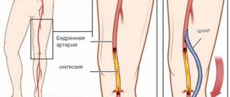
When using a contrast agent, it is injected into one of the veins or arteries on the thigh - depending on the type of drug and the purpose of the study. In this case, the ultrasound sensor remains on the patient in order to register all data and take measurements in a timely manner.
Preparation for echo-CG
As a rule, when performing one- and two-dimensional echocardiography, as well as Doppler echocardiography, there is no need for any special preparation. If a transesophageal study is prescribed, there are a number of limitations.
So, the last meal should be no later than six hours before the procedure. Drinking is also not recommended. Immediately before the procedure, dentures should be removed.
On the eve of a transesophageal examination, persons with a labile nervous system are recommended to take a mild sedative. After the procedure, the patient will need some time to recover, so you should not overload yourself with work until the end of the day. It is also necessary to refrain from driving.
Decoding the results
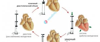
During the study, the doctor evaluates the linear dimensions and volumes of the heart chambers, local and general (ejection fraction) contractility and the speed of blood flow through the valves and in the vessels.
Basic indicators:
| Index | Meaning for women | Meaning for men | ||||||
| norm | minor violation | moderate impairment | significant violation | norm | minor violation | moderate impairment | significant violation | |
| Thickness of the interventricular septum | 6-9 mm | 10-12 mm | 13-15 mm | more than 16 mm | 6-10 mm | 11-13 mm | 14-16 mm | more than 17 mm |
| Thickness of the posterior wall of the left ventricle | 6-9 mm | 10-12 mm | 13-15 mm | more than 16 mm | 6-10 mm | 11-13 mm | 14-16 mm | more than 17 mm |
| Left ventricular myocardial mass (LVMM) | 66-150 g | 151-171 g | 172-192 g | more than 193 g | 66-150 g | 151-171 g | 172-192 g | more than 193 g |
| LVM index (a more significant indicator - takes into account the patient’s height) | 43-95 g/m2 | 96-108 g/m2 | 109-121 g/m2 | more than 122 g/m2 | 49-115 g/m2 | 116-131 g/m2 | 132-148 g/m2 | more than 149 g/m2 |
| Ejection fraction | more than 55% | 45-54% | 30-44% | less than 30% | more than 55% | 45-54% | 30-44% | less than 30% |
| Contractility | not violated | |||||||
In conclusion, the patient may notice the following terms:
- akinesis - absence of muscle/wall contraction;
- hypokinesis - minimal contraction;
- dyskinesis - asynchrony of wall contractions;
- hypertrophy - thickening;
- diastolic dysfunction - impaired relaxation of the heart muscle;
- systolic dysfunction - impaired myocardial contractility;
- dilatation - expansion of the cavity.
TERMINOLOGY AND DEFINITIONS
A summary of the terminology used in this echocardiographic technique and described in the text is given in Table 1.
Table 1. Speckle-tracking echocardiographic terminology
Strain Tension - Myocardial Strain
Strain rate - Myocardial strain rate
Longitudinal strain - Myocardial strain directed from the base to the apex of the heart
Radial strain Radial strain - Myocardial deformation directed radially to the center of the cavity of the left ventricle
Circumferential strain - Contraction of the left ventricle along the circular perimeter, observed in the short axis plane
Twisting - Net difference between mean apical and basal rotation in systole
Torsion (torsion) Torsion - Torsion of the LV normalized to the base-apex distance
Untwisting - Net difference between mean apical and basal rotation in diastole
Untwisting rate Untwisting rate
Bull's-eye - Topographic image warp value for all 17 segments
Post-systolic index — Percentage of post-systolic strain compared to maximum peak strain
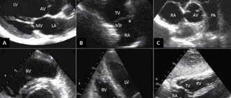
What does echocardiography show?
During the procedure, the doctor performs structural imaging and hemodynamic (Doppler) imaging.
Structural visualization includes:
- Visualization of the pericardium (eg, to exclude pericardial effusion);
- Visualization of the left or right ventricle and their cavities (to assess ventricular hypertrophy, wall motion abnormalities, and also to visualize blood clots);
- Valve imaging (mitral stenosis, aortic stenosis, mitral valve prolapse);
- Visualization of the great vessels (aortic dissection);
- Visualization of the atria and septa between the chambers of the heart (congenital heart disease, traumatic injuries).
Dopplerography:
- Visualization of blood flow through heart valves (valvular stenosis and regurgitation)
- Visualization of blood flow through the chambers of the heart (calculation of cardiac output, assessment of diastolic and systolic cardiac function)
Echocardiography for heart failure
- Makes it possible to determine the causes of the development of the chronic form (CHF)
- Left ventricular ejection fraction: reduced (systolic CHF);
- normal (diastolic CHF).
Echocardiography for cardiac arrhythmias
Important! Tachycardia makes it difficult to conduct research and accurately assess the results obtained.
Transthoracic echocardiography and emergency echocardiography are used.
Important parameters:
- Dimensions of the cavities of the heart, especially the atria
- Presence of blood clots
- Ejection fraction estimates are not always reliable
Echocardiography for assessing the condition of cancer patients
Heart damage in cancer patients:
- tumor intoxication;
- cardiotoxicity of chemotherapy drugs;
- cardiotoxicity of radiation therapy.
Indications for specialized echocardiography to determine cardiotoxicity:
- all patients with cancer before starting treatment;
- receiving chemotherapy or radiation therapy (after each course);
- have received chemotherapy or radiation therapy in the past (the frequency depends on the treatment regimen).
Longitudinal strain (stress) ― Longitudinal Strain
Longitudinal strain is the myocardial deformation directed from the base to the apex of the heart. During systole, the ventricular myocardial fibers shorten with a translational movement from the base to the apex. The subsequent reduction in the distance between individual nuclei is represented by negative trend curves (Fig. 1A).
By analyzing longitudinal strain in the 4-chamber, 2-chamber, and apical long-axis planes, both regional (relative to each of the 17 LV segments) and global strain values (global longitudinal strain) can be obtained. Global longitudinal strain has recently been validated as a quantitative measure to assess global LV function. The same measurements can be applied to speckle-tracking echocardiographic analysis of myocardial longitudinal strain of the left atrium and right ventricle (RV), obtaining peak longitudinal atrial strain and RV longitudinal strain, respectively.
Comparative characteristics of echocardiography with other methods
Echocardiography, despite its enormous benefits, remains a subjective method. Doctors take measurements and interpret some indicators differently. Sometimes ECHO-CG alone is not enough or it is impossible to carry out for some reason. Then other heart research techniques will come to the rescue:
- An electrocardiogram is the easiest way to detect heart problems. All medical and preventive institutions in the country and ambulances are equipped with ECG devices. An ECG reveals rhythm disturbances and gives a probable characteristic of local contractility (changes during myocardial infarction), but with ECHO-CG everything may look completely different.
- Chest x-ray allows you to determine the boundaries of the heart and detect cardiomegaly. To accurately diagnose the causes, visualization of the organ is necessary.
- Magnetic resonance imaging is used to clarify echocardiography data, study the morphology and functions of the heart chambers and valves. Now it is increasingly used for the diagnosis of non-coronary myocardial diseases and various cardiomyopathies.
- Contrast-enhanced computed tomography is very effective in examining the coronary arteries. Its cost is less than CAG (coronary angiography). It is also possible to determine all the sizes and volumes of the heart, but it will not allow you to see its work in real time.
- Coronary angiography (CAG) is the “gold standard” in the study of coronary arteries using a contrast agent with the identification of stenoses and their subsequent possible stenting (a method of expanding the lumen of the vessel). Its disadvantages are the very high cost and invasiveness of the procedure.
- Positron emission tomography is a rare study involving the administration of a radiopharmaceutical. Perfectly visualizes areas with insufficient blood supply (ischemia) of the myocardium
Echocardiography is the main and most accessible method of cardiac imaging, allowing for an accurate diagnosis, timely treatment, and evaluation of its effectiveness. Despite the subjectivity of the method, with constant training, advanced training, and exchange of experience with colleagues, a specialist increases his literacy in examination and minimizes the possibility of errors.
Echocardiography does not stand still. Attempts continue to supplement the method with new visualization capabilities, some of which allow the research to be carried out as objectively as possible and to minimize differences in interpretation between different specialists.
Sources:
- Saidova M.A. Transesophageal echocardiography: indications, technique. — Diseases of the heart and blood vessels, No. 4 2007.
- Marwick TH, trans. from English: Ph.D. Krikunova P.V. Recommendations for the use of echocardiography in hypertension in adults: a report from the European Association of Cardiovascular Imaging (EACVI) and the American Society of Echocardiography (ASE). Systemic hypertension. No. 2 2021.
- Ishak A Mansi, MD. Echocardiography. — Medscape, Jan 2014.
Twisting and Torsion
Until recently, assessment of LV torsion was only possible using MRI, but speckle-tracking echocardiography has now become a promising new tool for analyzing LV torsion. Left ventricular torsion is a component of normal systolic contraction that arises from the mutual rotation of the LV apex and base during systole and represents an important aspect of cardiac biomechanics. As an intrinsic physiological characteristic of the heart, quantitative assessment of left ventricular torsion during speckle-tracking echocardiography is based on analysis of the mutual rotation of the apex and base of the LV during systole. Left ventricular torsion is calculated as the net difference in mean rotation between apical and basal levels (Figure 2). Left ventricular torsion is defined as LV torsion normalized to the base-apex distance.
CONCLUSIONS
Speckle-tracking echocardiography is a new sophisticated echocardiographic technique that works with a standard 2-dimensional image, free of the limitations of Doppler technologies, and provides a comprehensive analysis of global and regional myocardial deformation in all spatial planes. In addition, speckle-tracking echocardiography allows one to evaluate LV rotational and torsional dynamics - parameters of LV function that, before the introduction of this technique, were analyzed exclusively using MRI.
Over the past 3 years, the evidence base has been growing and demonstrates the good feasibility, reproducibility and accuracy of speckle-tracking echocardiography in a variety of clinical applications. However, there is still no published prospective clinical trial to evaluate this method in large populations. The recently developed 3D speckle-tracking technique has shown promising preliminary results in the evaluation of 3D image data. This is another technology advancement that is expected to provide more extensive and detailed analysis of cardiac dynamics, bringing echocardiography closer to the most advanced imaging modality, while maintaining the ability to perform it at the bedside.
rh.org.ru
- Views: 16051
- Comments: